Bolognia 5e
·
Dermatoloji Çalışma Paneli
▤
Anasayfa
📖
Özetler
🃏
Flashcard
❓
Quiz
🖼
Atlas
◐
Ch 14
Allerjik Kontakt Dermatit
Vol 1 · sayfa 246 · §3
← Ch 12
Ch 18 →
📖
Özet
🃏
Flashcard
❓
Quiz
🖼
Atlas
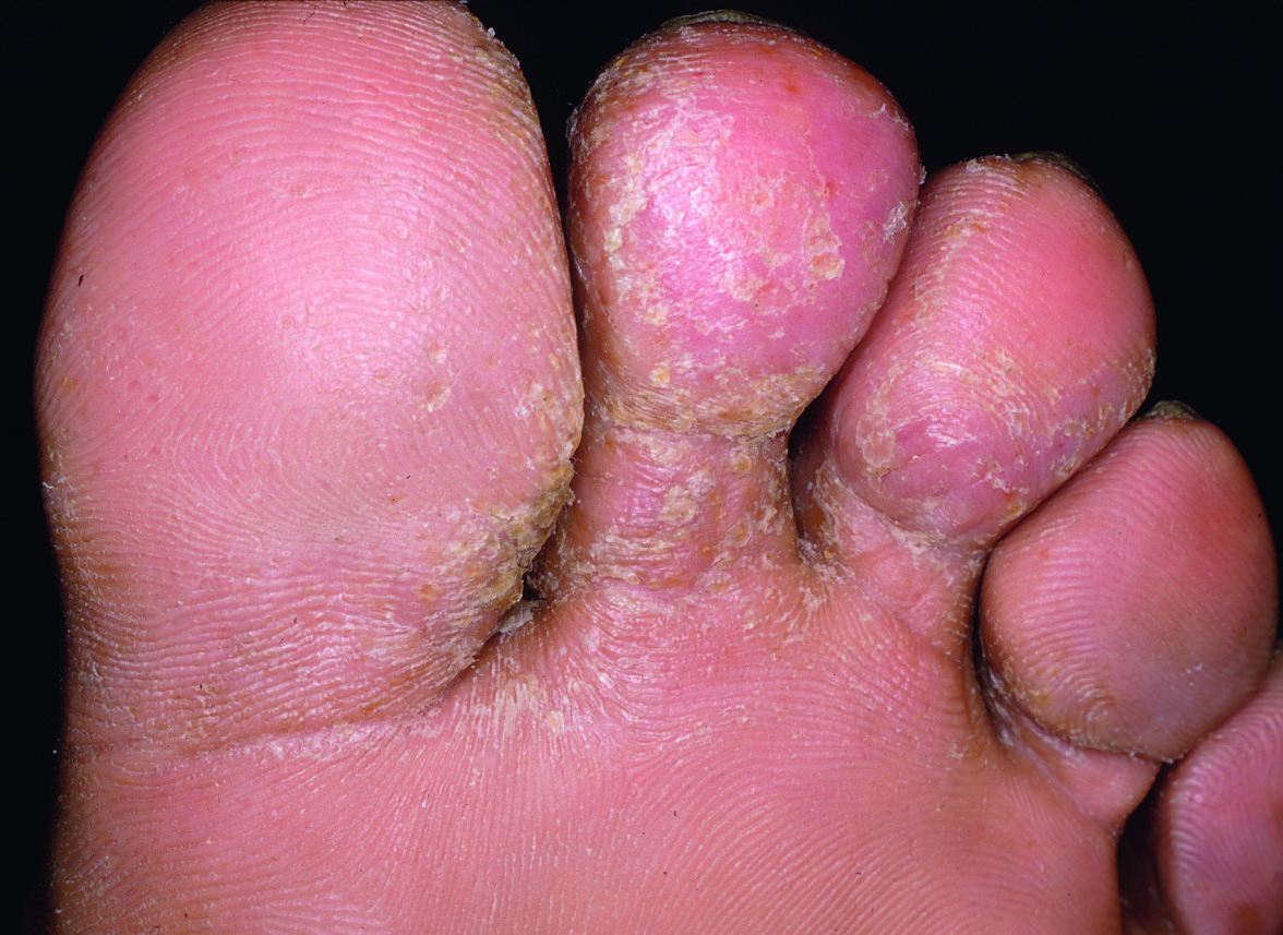
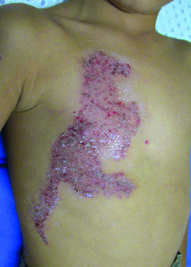
Fig.14.1 A
s.328
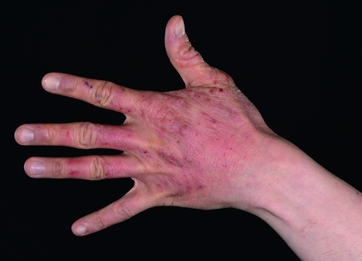
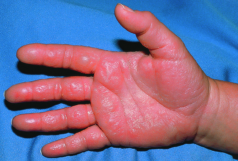
Fig.14.1 B
s.328
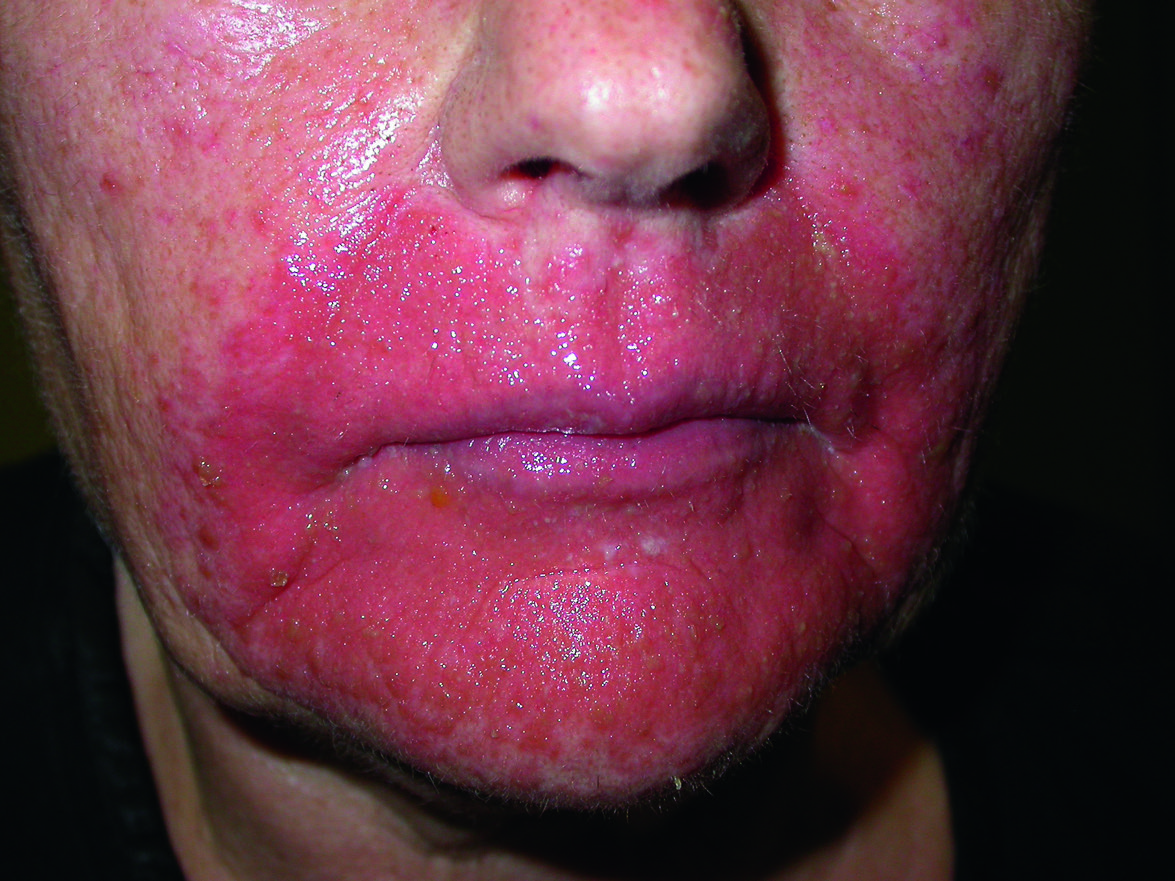
Fig.14.2
s.329
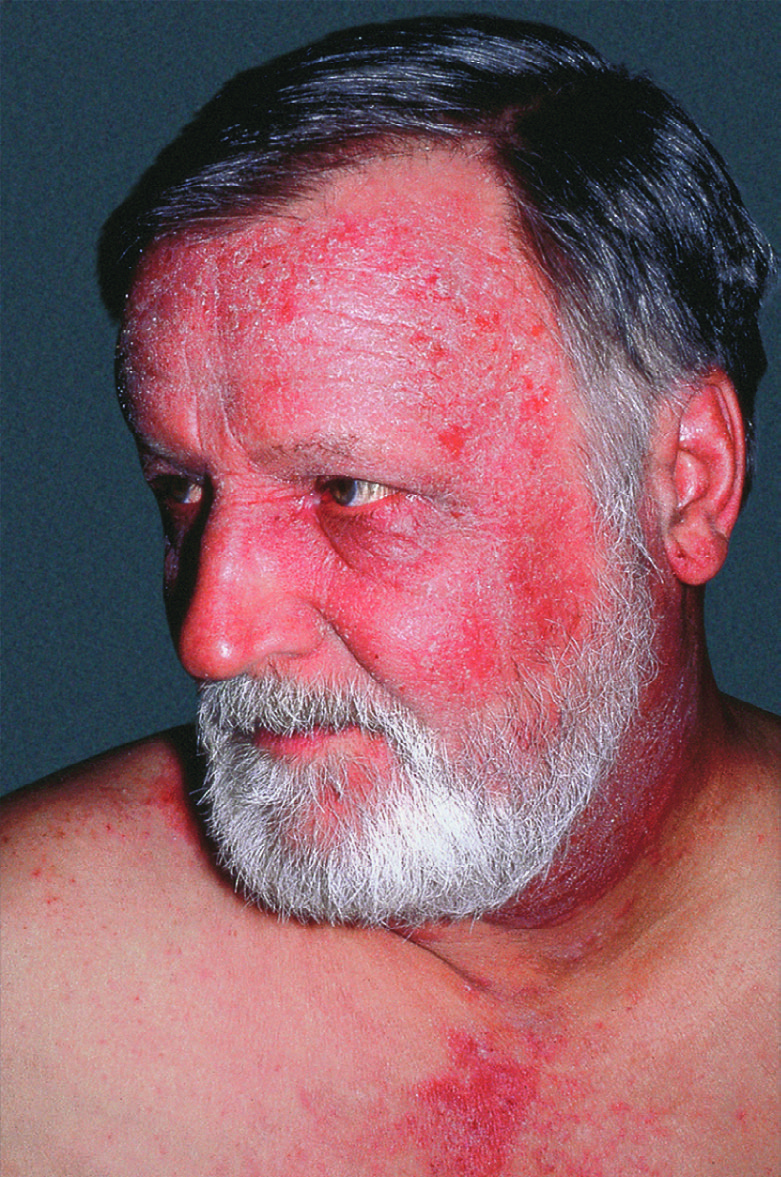
Fig.14.3
s.329
Fig.14.4 A
s.329
Fig.14.4 B
s.329
Fig.14.5 A
s.330
Fig.14.5 B
s.330
Fig.14.6 A
s.330
Fig.14.6 B
s.330
Fig.14.7
s.330
Fig.14.8 A
s.330
Fig.14.8 B
s.330
Fig.14.10 A
s.332
Fig.14.10 B
s.332
Fig.14.12
s.336
Fig.14.14
s.337
Fig.14.15 A
s.338
Fig.14.15 B
s.338
Fig.14.17
s.343
Fig.14.19
s.345
Fig.14.21 A
s.346
Fig.14.21 B
s.346
Fig.14.21 C
s.346
✕ Kapat