Bolognia 5e·Dermatoloji Çalışma Paneli
Ch 18

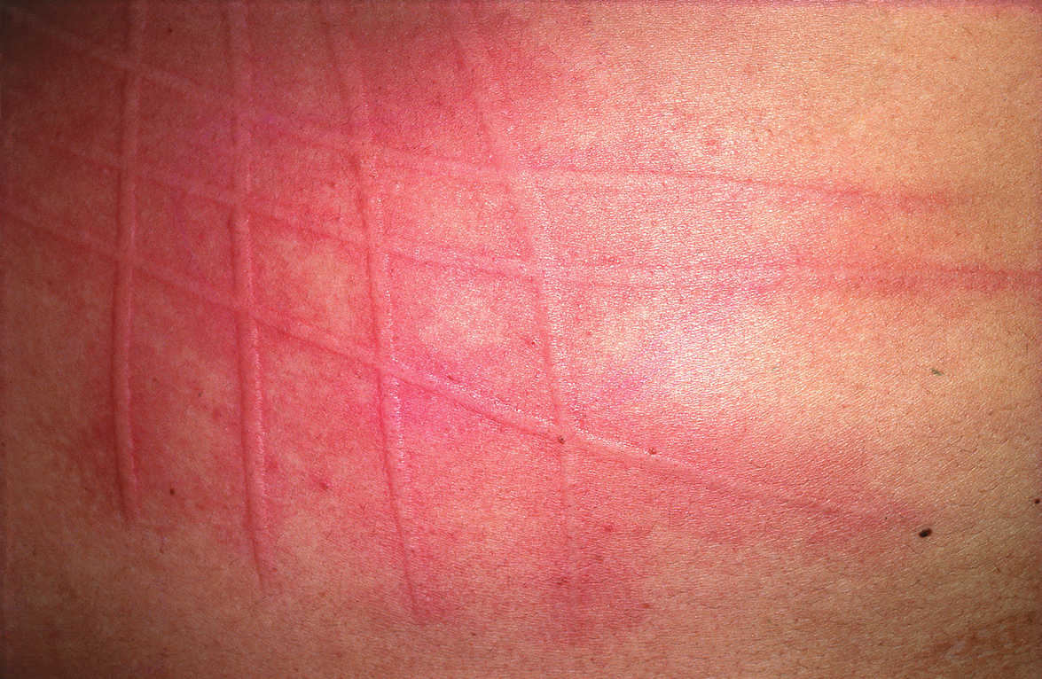
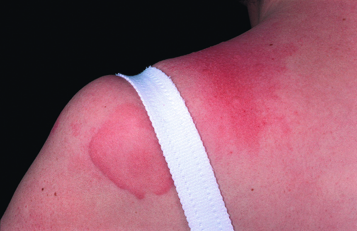
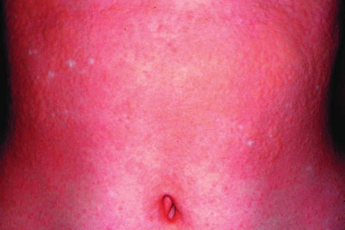
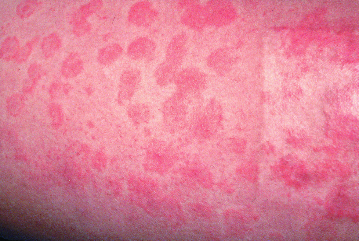
Ürtiker ve Anjiyoödem

Vol 1 · sayfa 309 · §4
📖 Özet🃏 Flashcard Quiz🖼 Atlas
Fig. 18.9 Symptomatic immediate dermographism. Itchy wheals appear very soon after scratching. Courtesy Jean L. Bolognia, MD.
Fig.18.9s.408
Fig. 18.10 Delayed pressure urticaria.
Fig.18.10s.409
Fig. 18.11 Cold urticaria. Wheals developed on the forearm after placement of an ice cube for 10 minutes, followed by rewarming. Courtesy Thomas Schwarz, MD.
Fig.18.11s.409
Fig. 18.12 Cholinergic urticaria on the trunk after hot bath provocation.
Fig.18.12s.410
Fig. 18.13 Urticarial vasculitis. Lesions look like spontaneous urticaria but last longer and may bruise. An incidental finding is a skin graft donor site.
Fig.18.13s.411
Fig. 18.14 Spontaneous urticaria – histologic findings. A sparse, mainly perivascular inflammatory infiltrate and scant edema within the superficial and mid dermis. Note several neutrophils and a few eosinophils (inset). Courtesy Lorenzo Cerroni, MD.
Fig.18.14s.412
Fig. 18.15 Urticaria in a young child. This clinical presentation of urticaria is sometimes misdiagnosed as erythema multiforme because of the dusky centers, with some clinicians referring to it as urticaria multiforme. As lesions expand, central clearing can occur, which is not seen in erythema multiforme. Courtesy Luis Requena, MD.
Fig.18.15s.412
Fig. 18.17 Autologous serum skin test (ASST). At 30 minutes, a red wheal response is present at the site of an intradermal injection of autologous serum, but not at that of a normal saline control. For the test to be positive, the wheal response at the serum injection site must be at least 1.5 mm greater in diameter than the control site. This criterion is a reasonably sensitive and specific screening test for functional autoanti- bodies as detected by the basophil release assay. Spontaneous wheals are also present at the venipuncture site and above the serum skin test.
Fig.18.17s.413