Bolognia 5e
·
Dermatoloji Çalışma Paneli
▤
Anasayfa
📖
Özetler
🃏
Flashcard
❓
Quiz
🖼
Atlas
◐
Ch 42
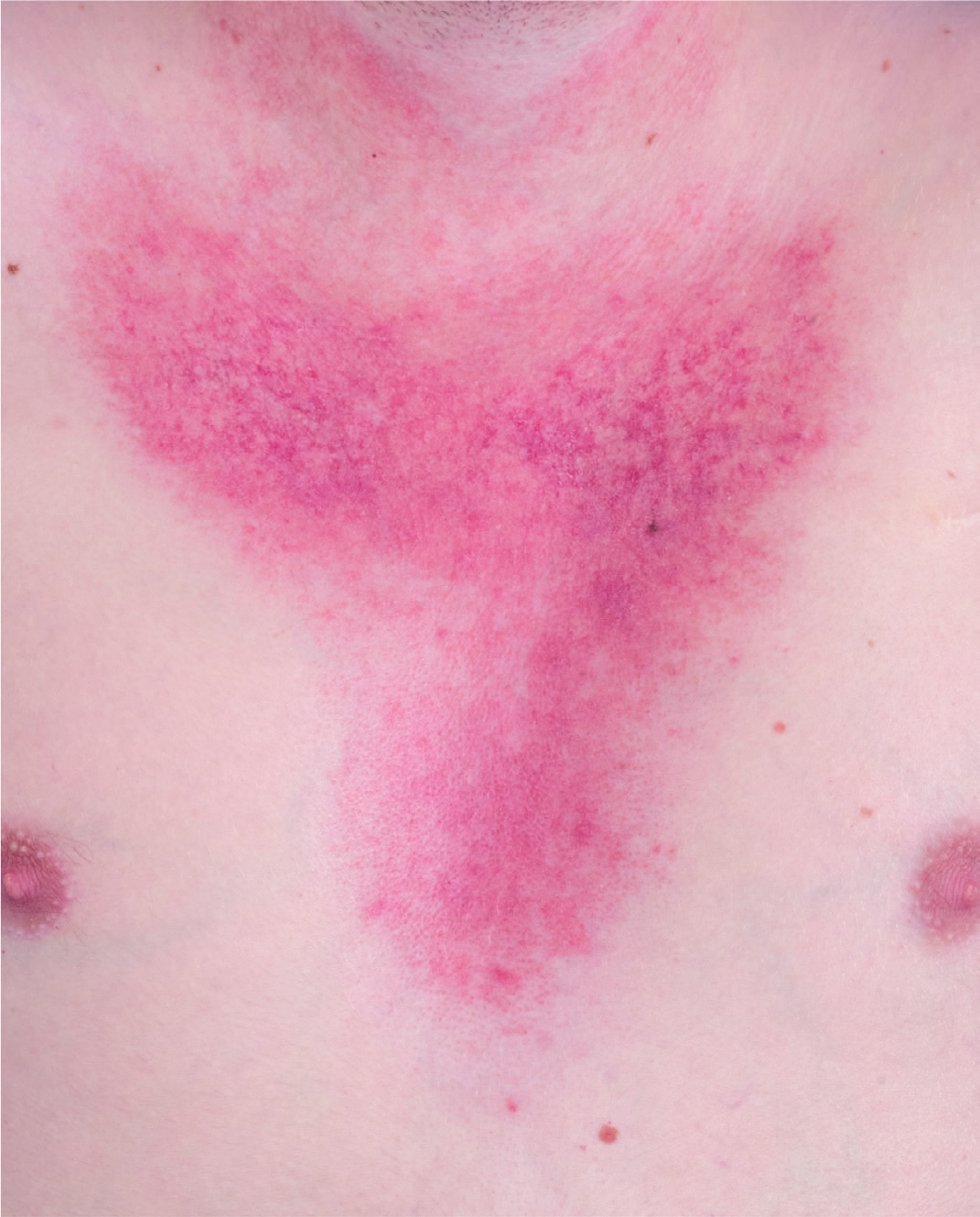
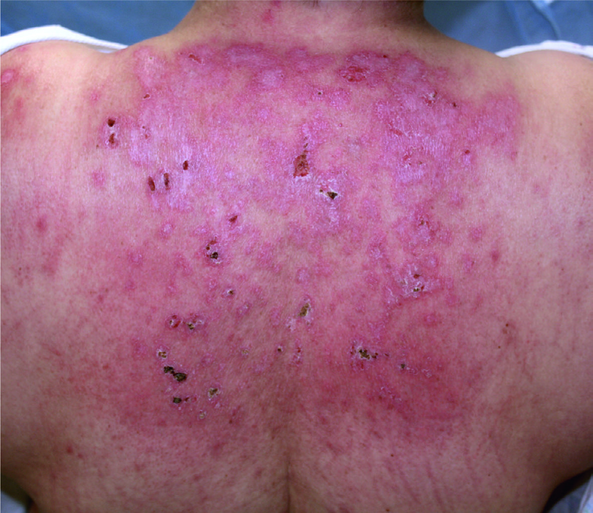
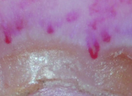
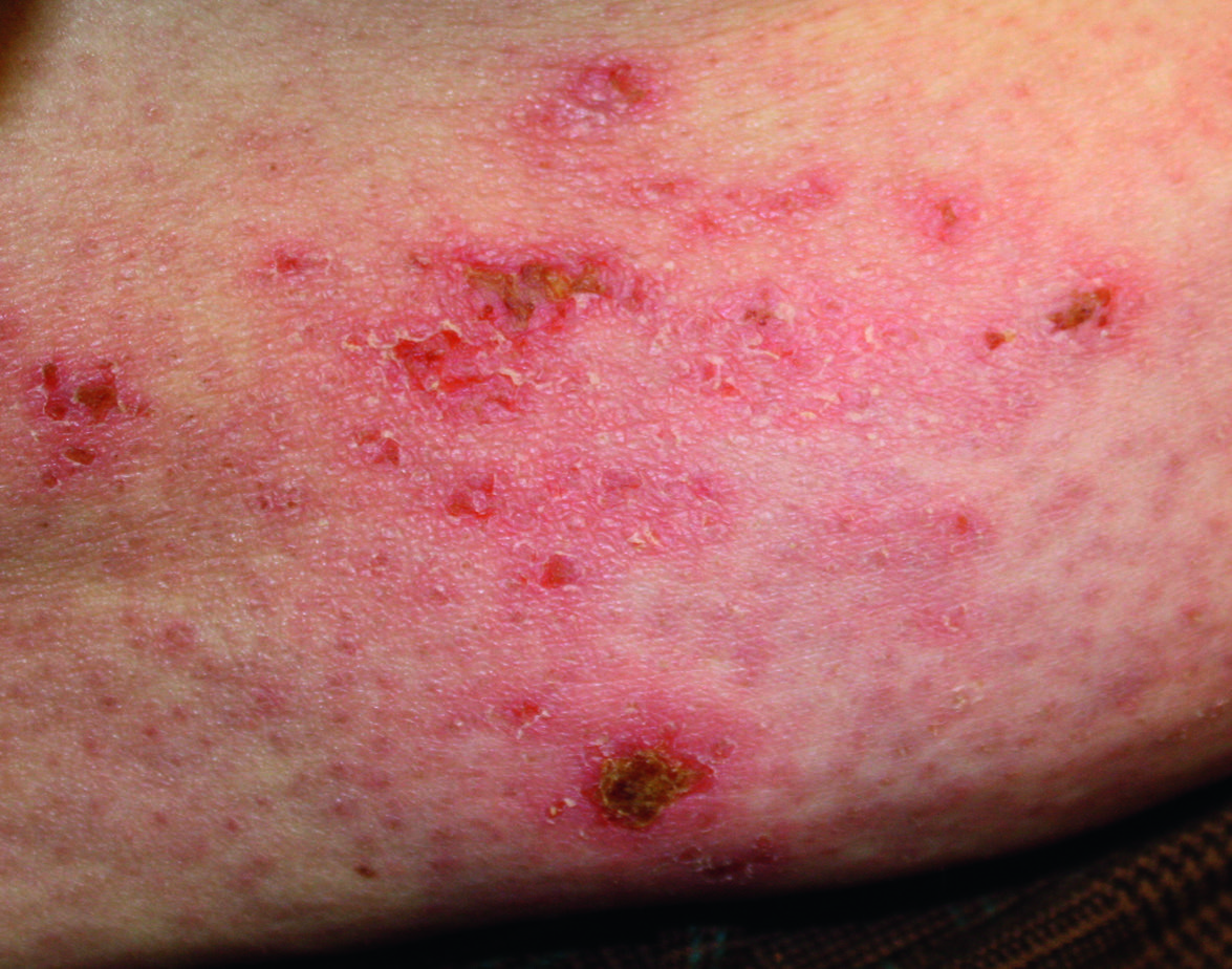
Dermatomiyozit
Vol 1 · sayfa 689 · §7
← Ch 41
Ch 43 →
📖
Özet
🃏
Flashcard
❓
Quiz
🖼
Atlas
Fig.42.2 A
s.865
Fig.42.2 B
s.865
Fig.42.3
s.865
Fig.42.4 A
s.866
Fig.42.4 B
s.866
Fig.42.4 C
s.866
Fig.42.4 D
s.866
Fig.42.5 A
s.866
Fig.42.5 B
s.866
Fig.42.6 A
s.867
Fig.42.6 B
s.867
Fig.42.6 C
s.867
Fig.42.6 D
s.867
Fig.42.7
s.867
Fig.42.8
s.867
Fig.42.9
s.869
Fig.42.10 A
s.868
Fig.42.10 B
s.868
Fig.42.11C
s.869
Fig.42.11
s.869
Fig.42.14
s.871
✕ Kapat