Bolognia 5e
·
Dermatoloji Çalışma Paneli
▤
Anasayfa
📖
Özetler
🃏
Flashcard
❓
Quiz
🖼
Atlas
◐
Ch 84
İnfestasyonlar
Vol 2 · sayfa 1519 · §12
← Ch 82
Ch 87 →
📖
Özet
🃏
Flashcard
❓
Quiz
🖼
Atlas
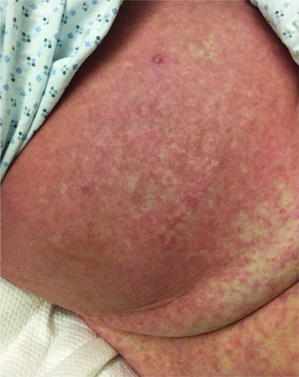
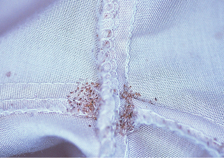
Fig.84.4
s.1841
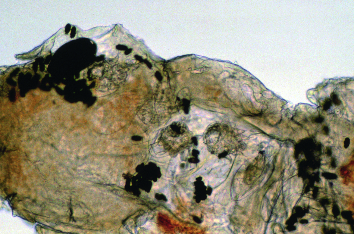
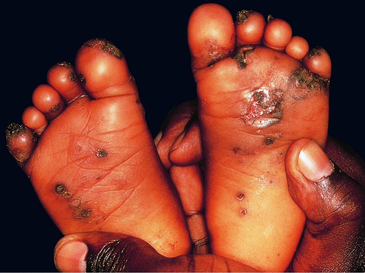
Fig.84.5
s.1841
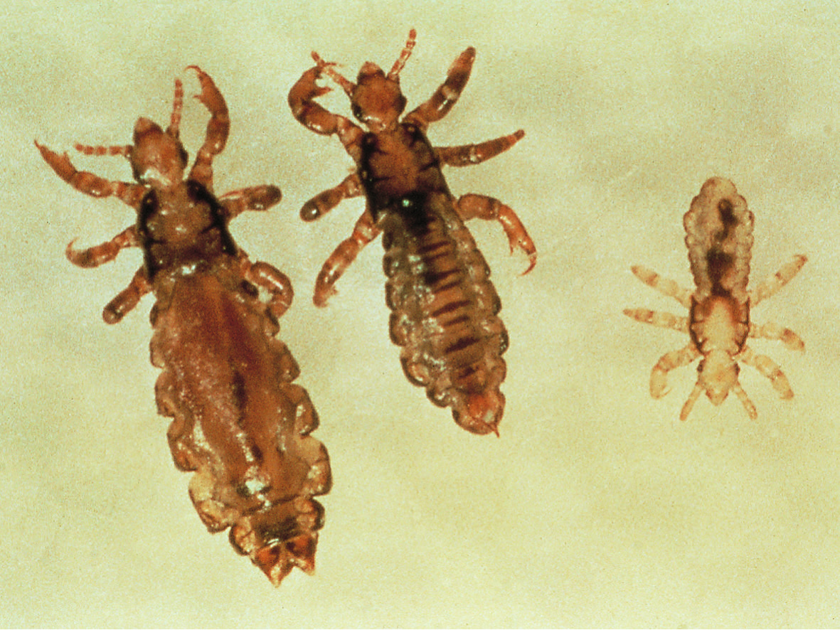
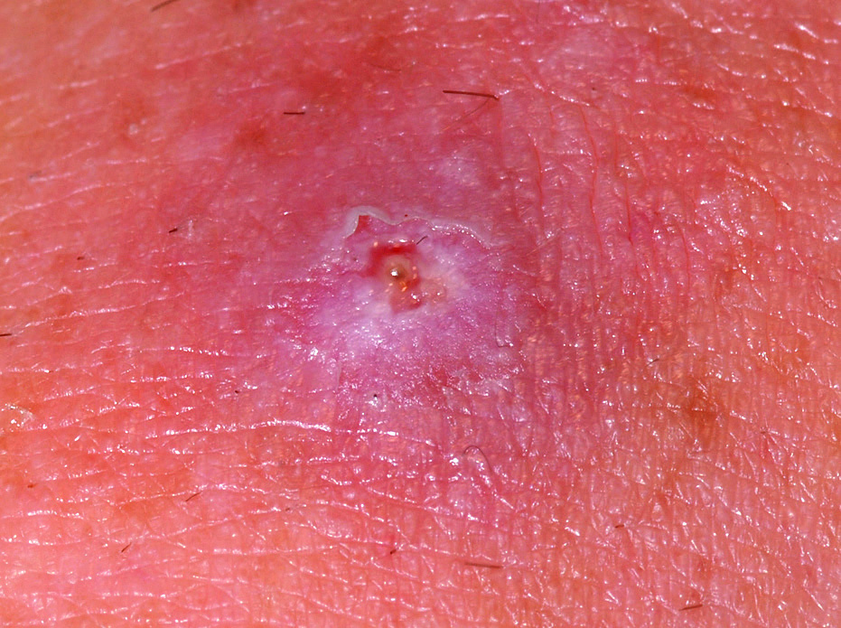
Fig.84.7
s.1843
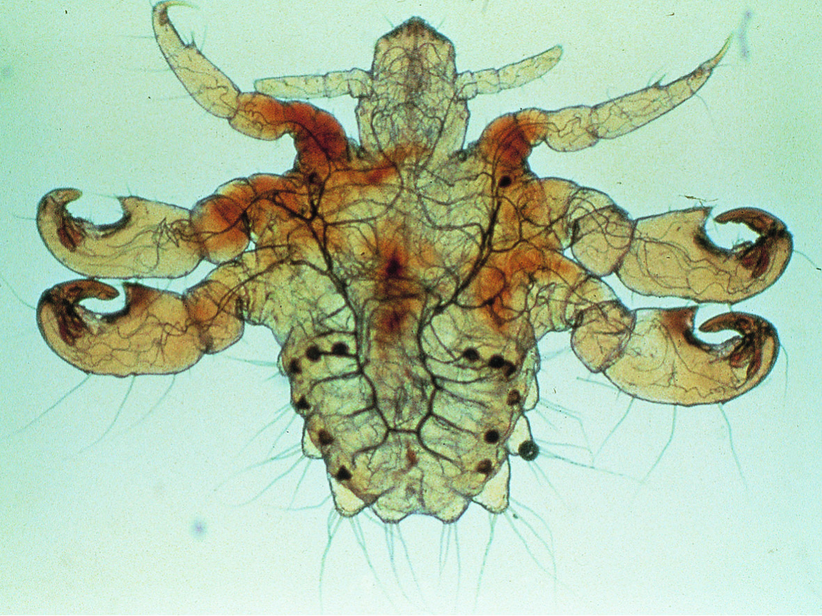
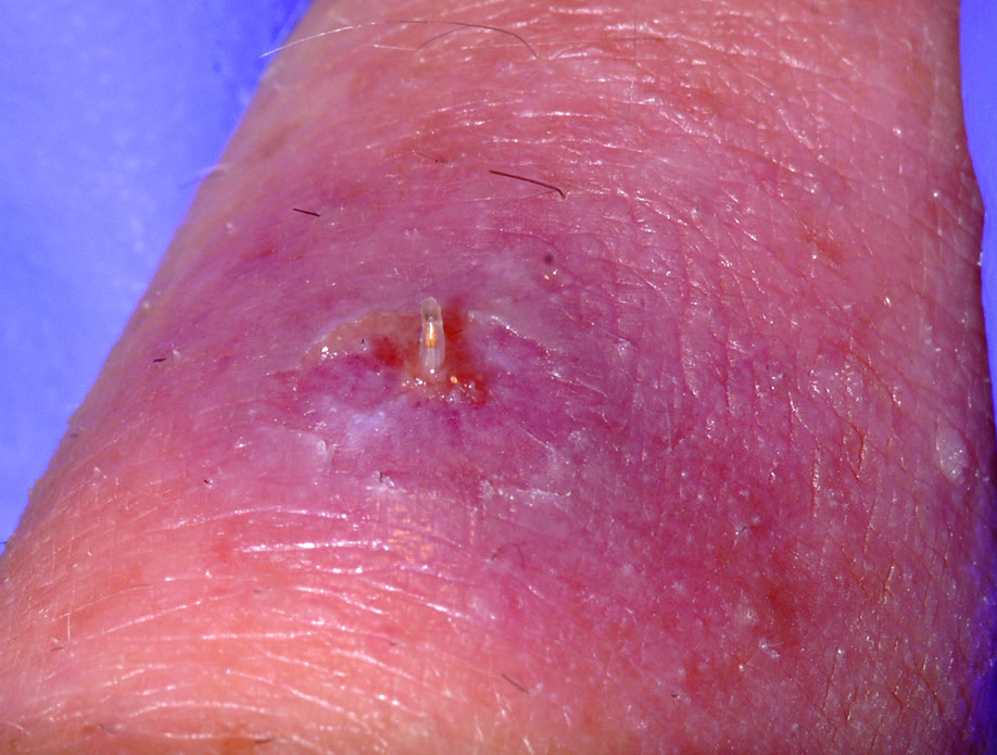
Fig.84.9 A
s.1844
Fig.84.9 B
s.1844
Fig.84.10
s.1847
Fig.84.11 A
s.1847
Fig.84.11 B
s.1847
Fig.84.12
s.1848
Fig.84.13
s.1849
Fig.84.14 A
s.1850
Fig.84.14 B
s.1850
Fig.84.15
s.1850
✕ Kapat