Bolognia 5e
·
Dermatoloji Çalışma Paneli
▤
Anasayfa
📖
Özetler
🃏
Flashcard
❓
Quiz
🖼
Atlas
◐
Ch 93
Non-infeksiyöz Granülomalar
Vol 2 · sayfa 1660 · §14
← Ch 87
Ch 108 →
📖
Özet
🃏
Flashcard
❓
Quiz
🖼
Atlas
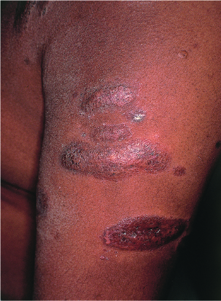
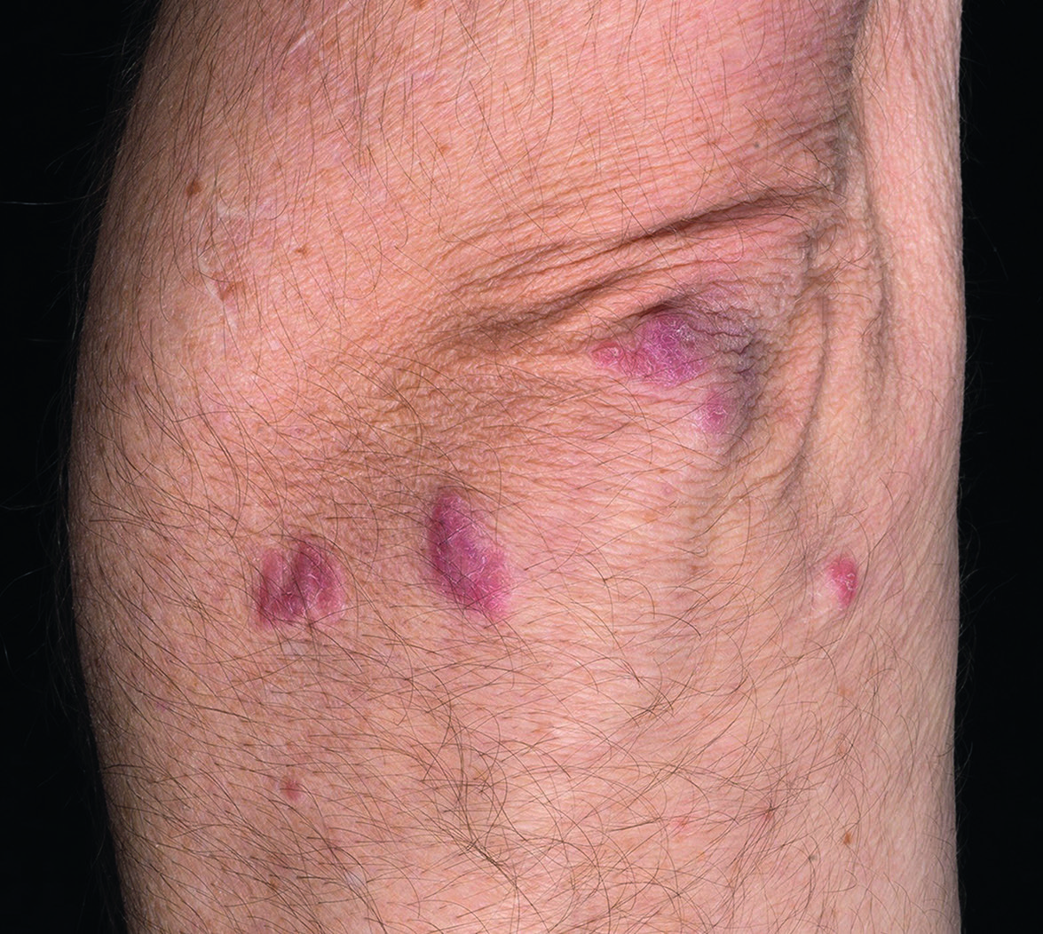
Fig.93.2 A
s.2005
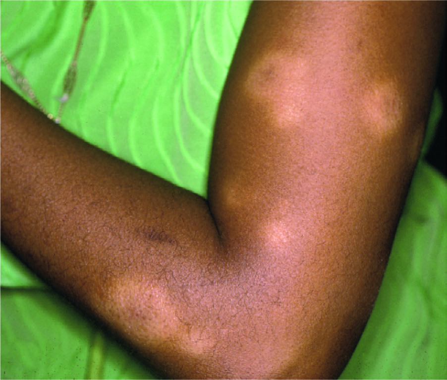
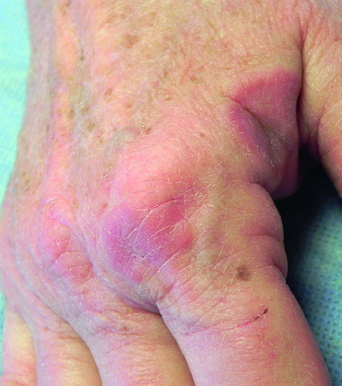
Fig.93.2 B
s.2005
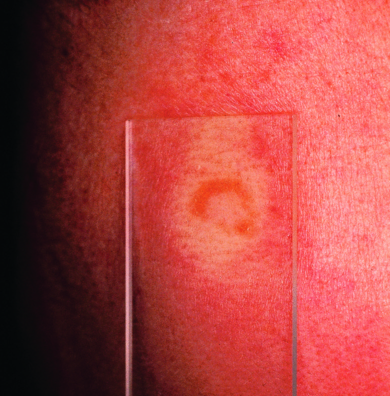
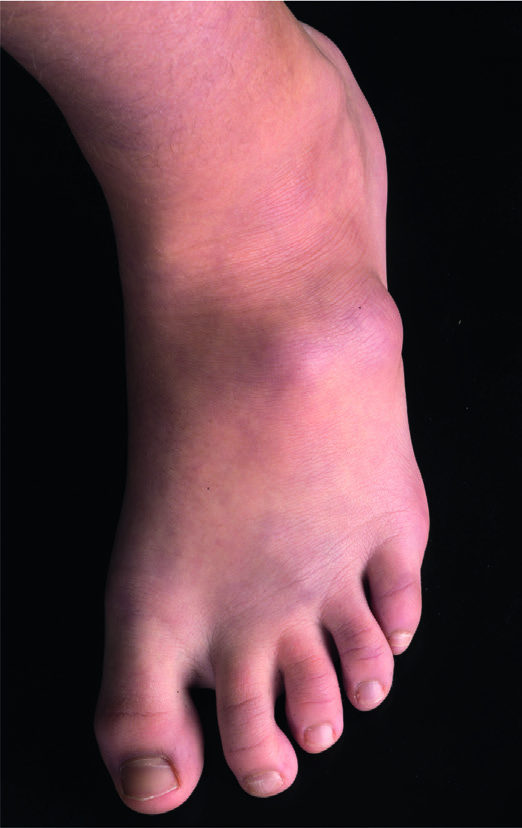
Fig.93.2 C
s.2005
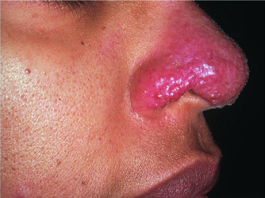
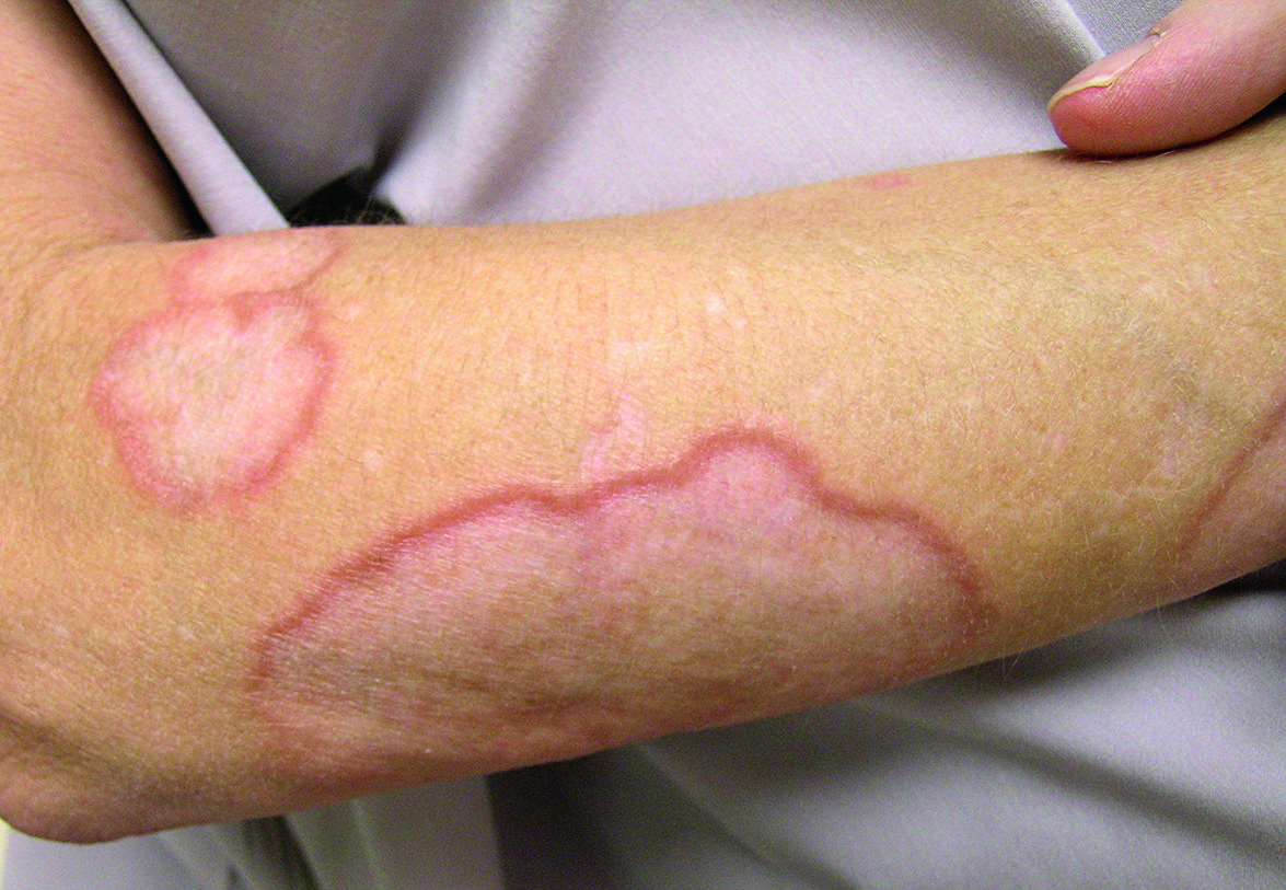
Fig.93.3 A
s.2006
Fig.93.3 B
s.2006
Fig.93.3 C
s.2006
Fig.93.4
s.2006
Fig.93.5
s.2006
Fig.93.8
s.2008
Fig.93.9 A
s.2011
Fig.93.9 B
s.2011
Fig.93.9 C
s.2011
Fig.93.9 D
s.2011
Fig.93.10 A
s.2012
Fig.93.10 B
s.2012
Fig.93.10 C
s.2012
Fig.93.11 A
s.2013
Fig.93.11 B
s.2013
Fig.93.12
s.2013
Fig.93.13 A
s.2013
Fig.93.13 B
s.2013
Fig.93.15 A
s.2016
Fig.93.15 B
s.2016
Fig.93.15 C
s.2016
Fig.93.15 D
s.2016
Fig.93.15 E
s.2016
Fig.93.17 A
s.2017
Fig.93.17 B
s.2017
Fig.93.17 C
s.2017
Fig.93.17 D
s.2017
Fig.93.17 E
s.2017
Fig.93.17 F
s.2017
Fig.93.18 A
s.2019
Fig.93.18 B
s.2019
Fig.93.18 C
s.2019
✕ Kapat