Bolognia 5e
·
Dermatoloji Çalışma Paneli
▤
Anasayfa
📖
Özetler
🃏
Flashcard
❓
Quiz
🖼
Atlas
◐
Ch 108
AK, BCC ve SCC
Vol 2 · sayfa 1888 · §18
← Ch 93
Ch 113 →
📖
Özet
🃏
Flashcard
❓
Quiz
🖼
Atlas
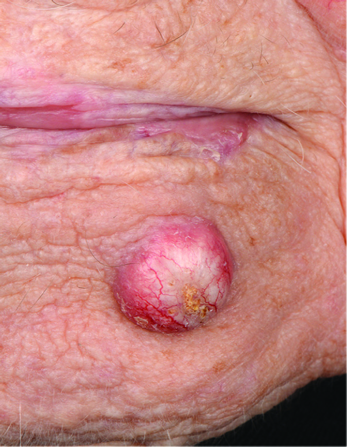
Fig.108.1 A
s.241
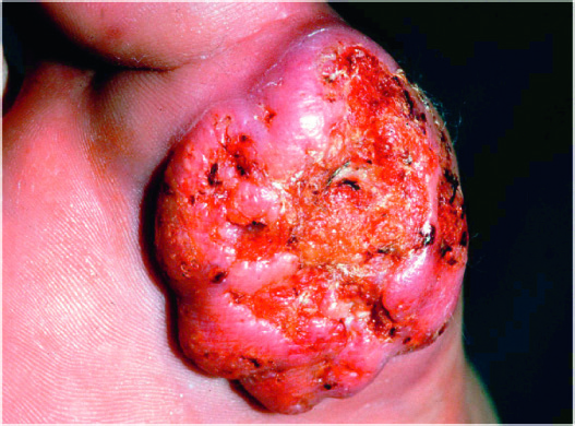
Fig.108.1 B
s.241
Fig.108.2 A
s.242
Fig.108.2 B
s.242
Fig.108.2 C
s.242
Fig.108.3 A
s.243
Fig.108.3 B
s.243
Fig.108.3 C
s.243
Fig.108.4 A
s.243
Fig.108.4 B
s.243
Fig.108.5 A
s.243
Fig.108.5 B
s.243
Fig.108.5 C
s.243
Fig.108.5 D
s.243
Fig.108.6 A
s.244
Fig.108.6 B
s.244
Fig.108.6 C
s.244
Fig.108.6 D
s.244
Fig.108.6 E
s.244
Fig.108.6 F
s.244
Fig.108.9 A
s.246
Fig.108.9 B
s.246
Fig.108.9 C
s.246
Fig.108.9 D
s.246
Fig.108.9 E
s.246
Fig.108.10A,B
s.246
Fig.108.10 A
s.247
Fig.108.10 B
s.247
Fig.108.10 C
s.247
Fig.108.10 D
s.247
Fig.108.17 A
s.250
Fig.108.17 B
s.250
Fig.108.17 C
s.250
Fig.108.17 D
s.250
Fig.108.17 E
s.250
Fig.108.17 F
s.250
Fig.108.17 G
s.250
Fig.108.18 A
s.251
Fig.108.18 B
s.251
Fig.108.18 C
s.251
Fig.108.19 A
s.251
Fig.108.19 B
s.251
Fig.108.19 C
s.251
Fig.108.19 D
s.251
Fig.108.23 A
s.257
Fig.108.23 B
s.257
✕ Kapat