Bolognia 5e
·
Dermatoloji Çalışma Paneli
▤
Anasayfa
📖
Özetler
🃏
Flashcard
❓
Quiz
🖼
Atlas
◐
Ch 20
Eritema Multiforme / SJS / TEN
Vol 1 · sayfa 339 · §4
← Ch 18
Ch 21 →
📖
Özet
🃏
Flashcard
❓
Quiz
🖼
Atlas
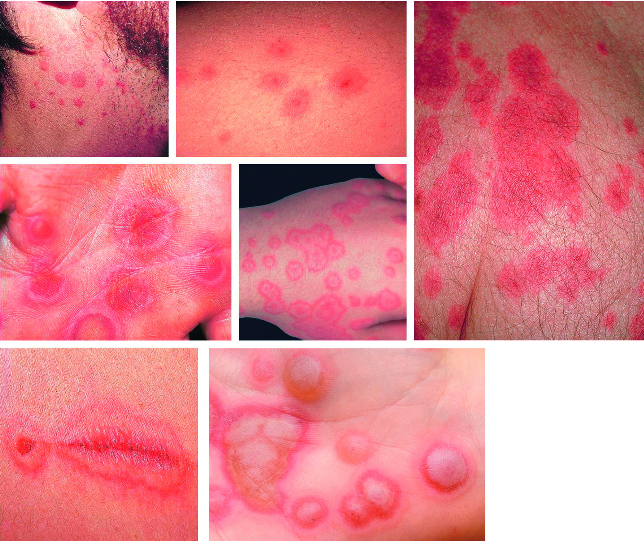
Fig.20.1
s.438
Fig.20.2
s.439
Fig.20.3 A
s.439
Fig.20.3 B
s.439
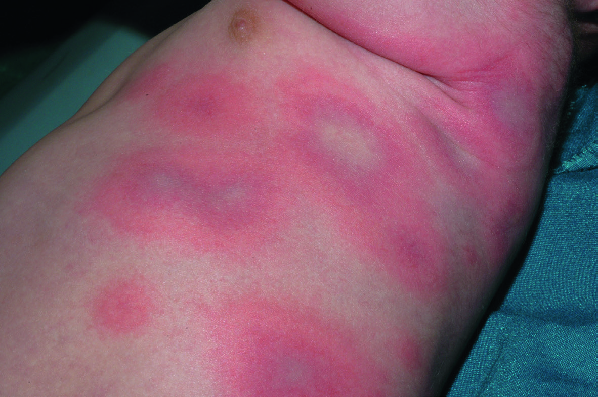
Fig.20.5 A
s.440
Fig.20.5 B
s.440
Fig.20.5 C
s.440
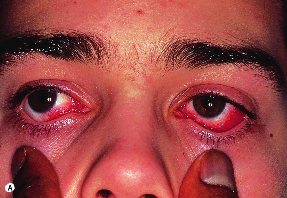
Fig.20.7 A
s.445
Fig.20.7 B
s.445
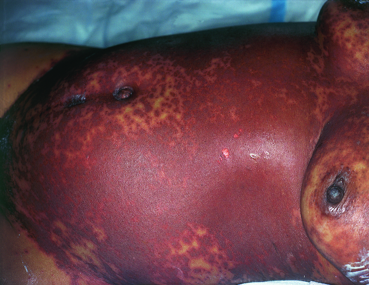
Fig.20.8
s.445
Fig.20.9 A
s.446
Fig.20.9 B
s.446
Fig.20.9 C
s.446
Fig.20.12 A
s.447
Fig.20.12 B
s.447
Fig.20.12 C
s.447
Fig.20.14 A
s.448
Fig.20.14 B
s.448
✕ Kapat