Bolognia 5e
·
Dermatoloji Çalışma Paneli
▤
Anasayfa
📖
Özetler
🃏
Flashcard
❓
Quiz
🖼
Atlas
◐
Ch 21
İlaç Reaksiyonları
Vol 1 · sayfa 355 · §4
← Ch 20
Ch 24 →
📖
Özet
🃏
Flashcard
❓
Quiz
🖼
Atlas
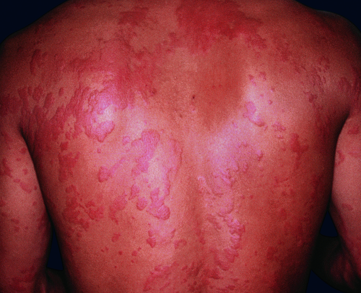
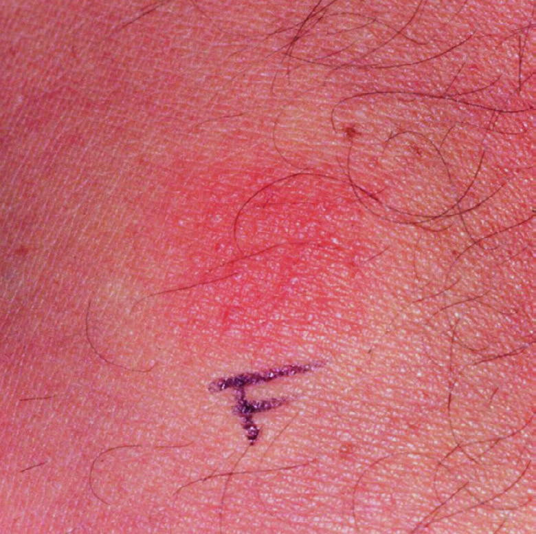
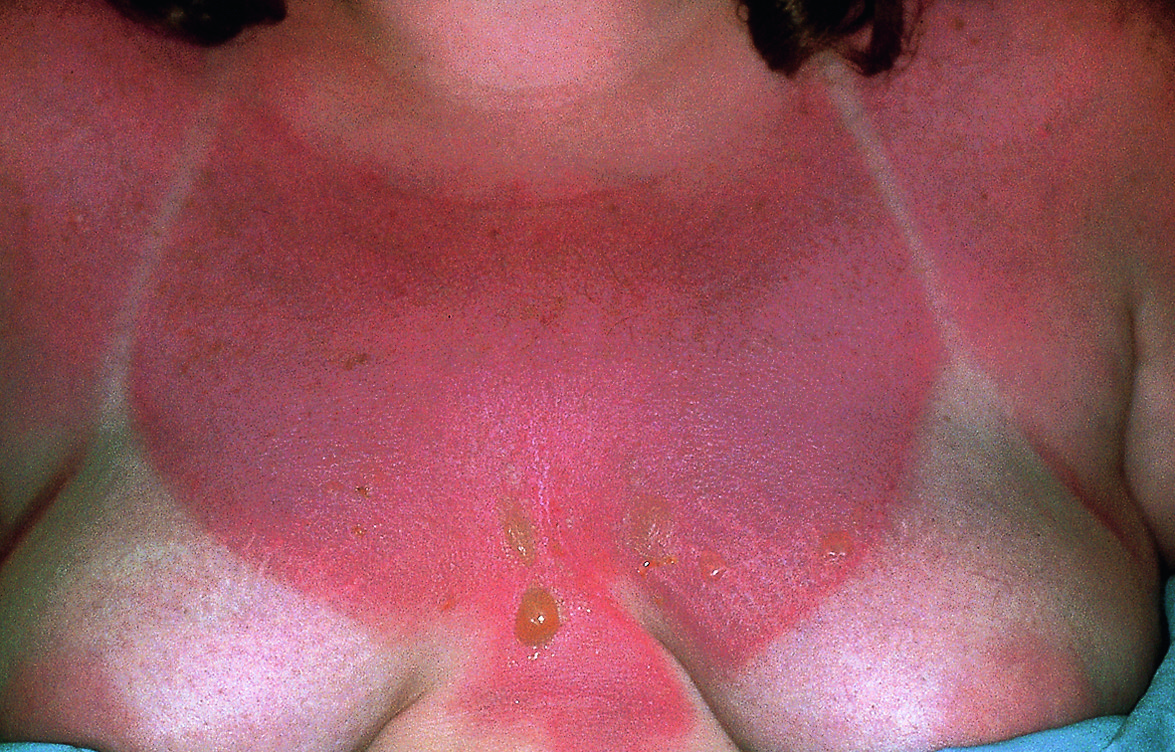
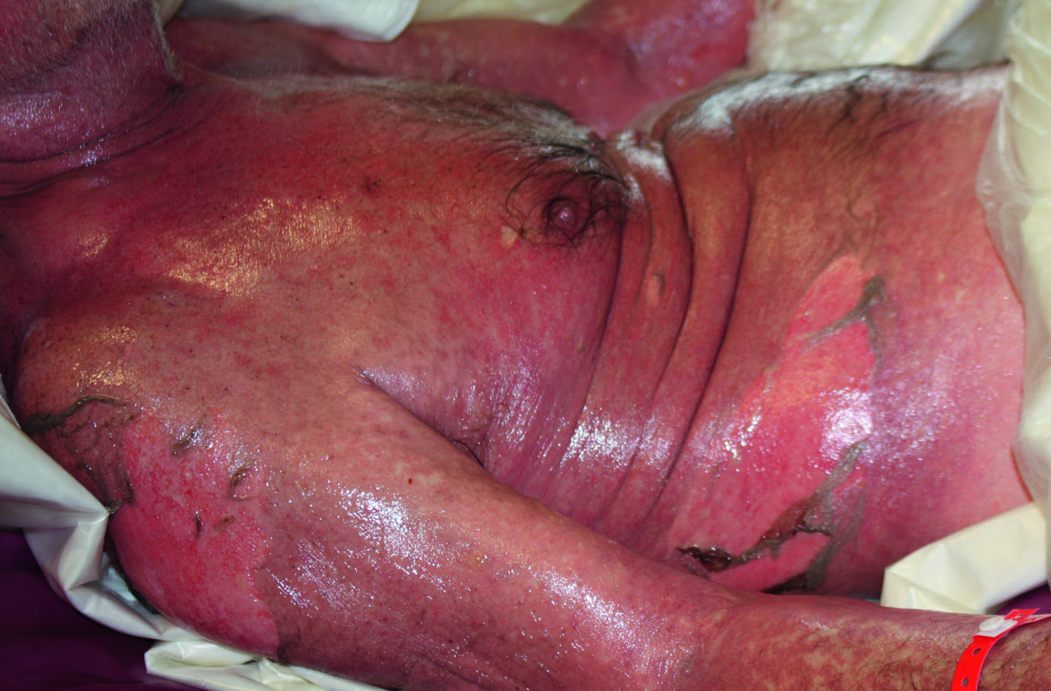
Fig.21.1 A
s.457
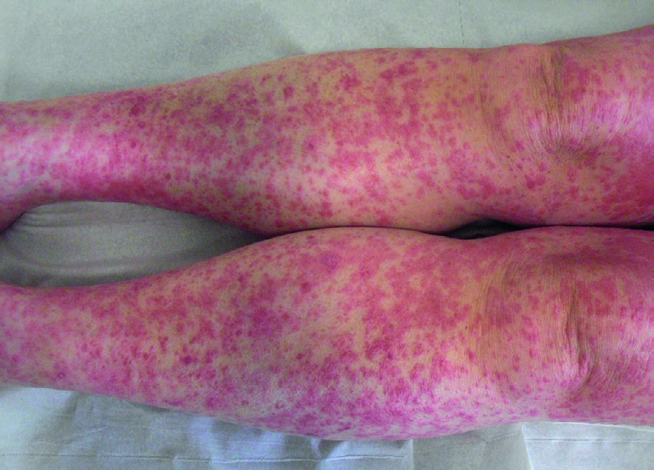
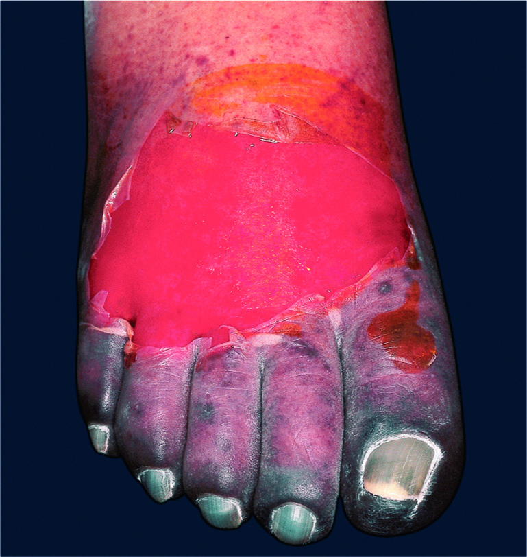
Fig.21.1 B
s.457
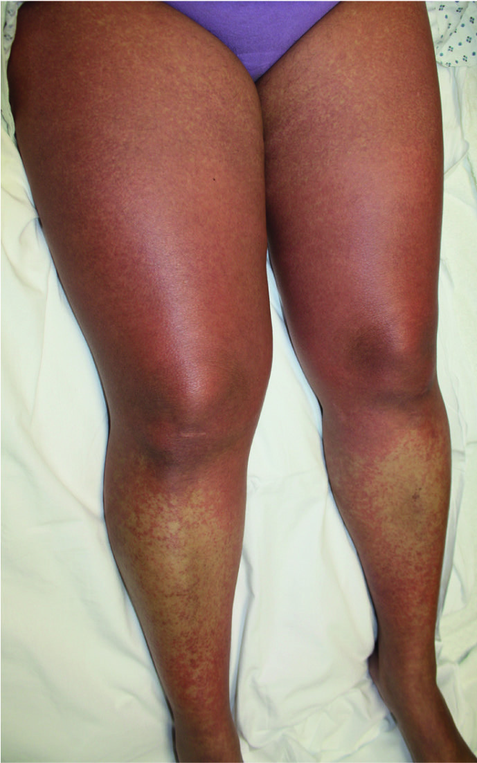
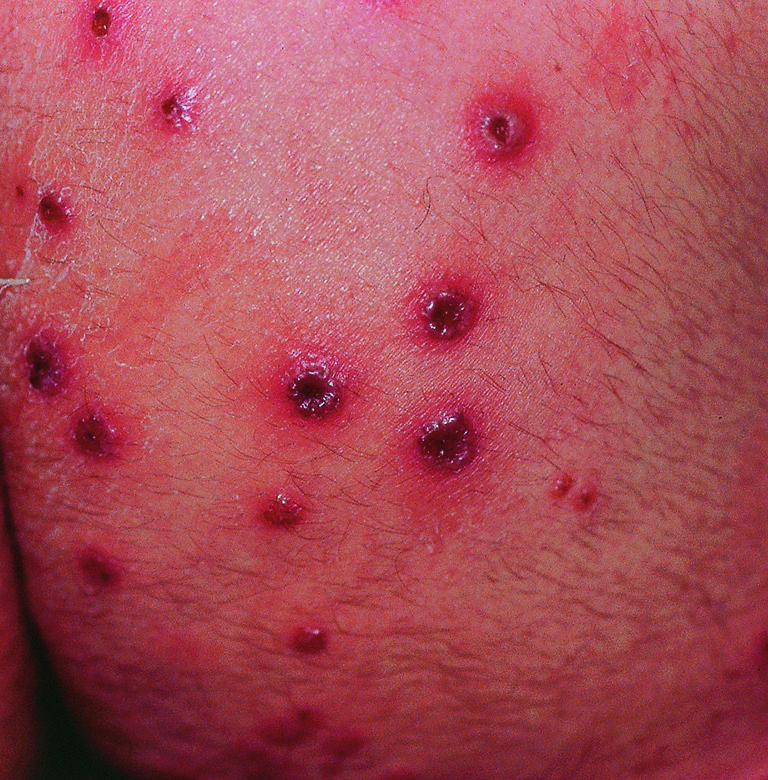
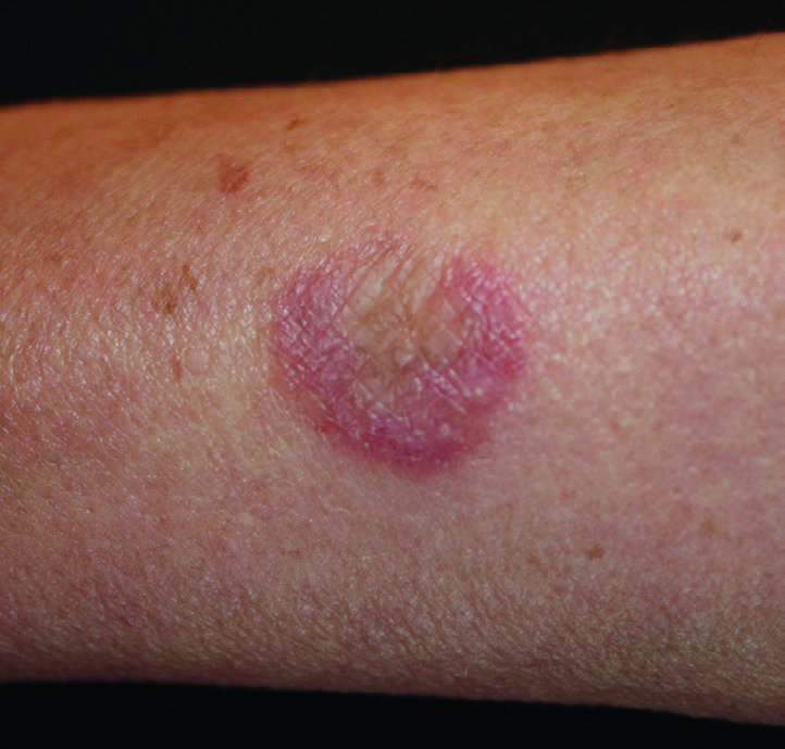
Fig.21.2
s.460
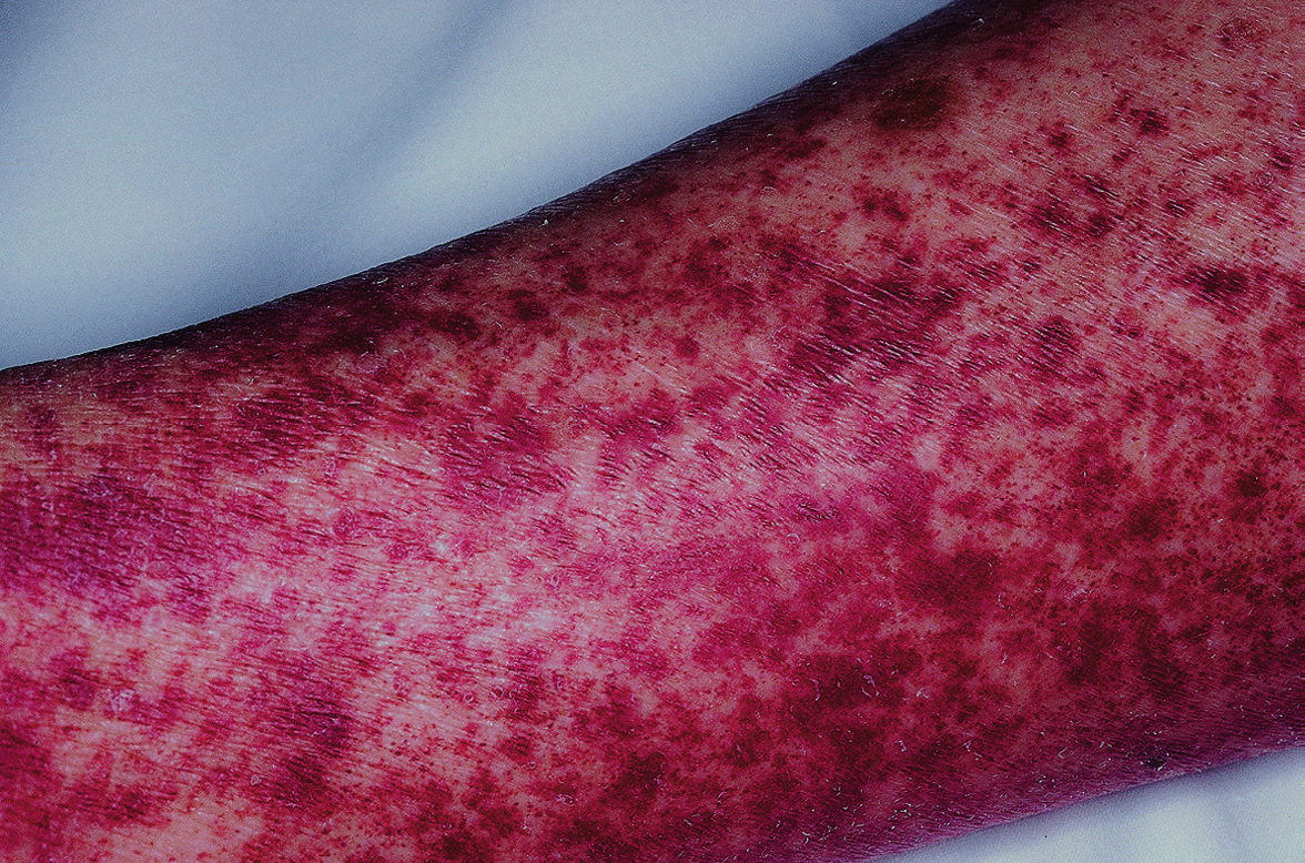
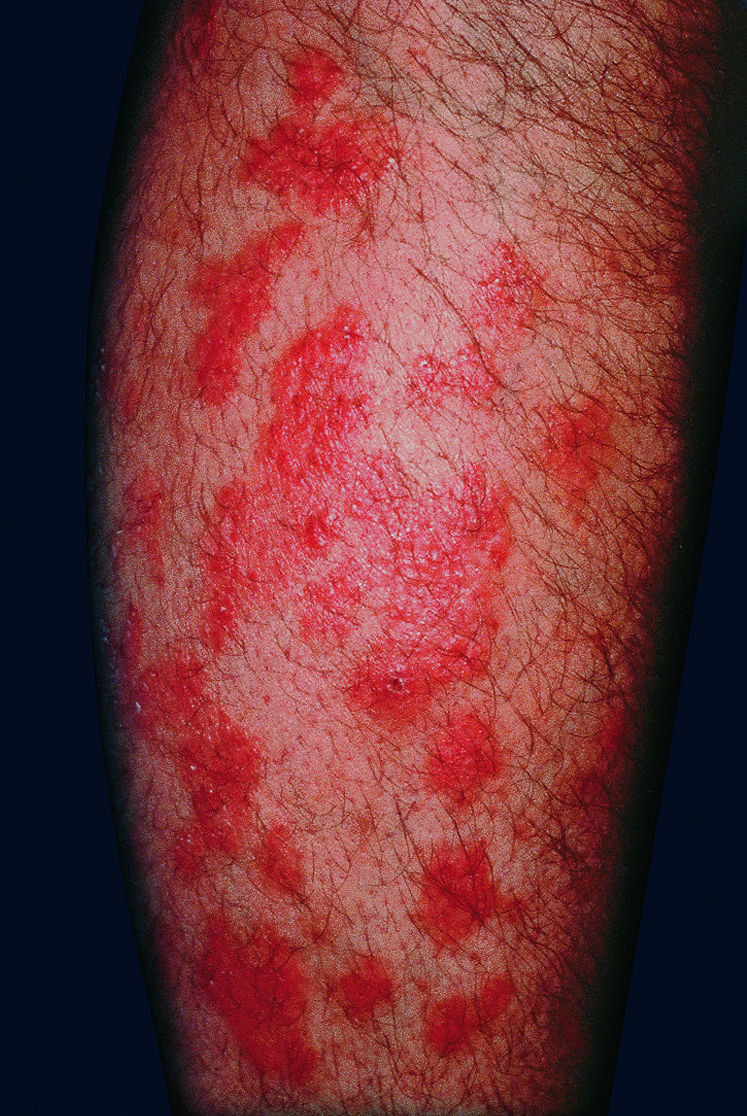
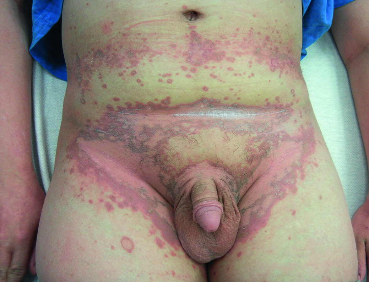
Fig.21.3 A
s.461
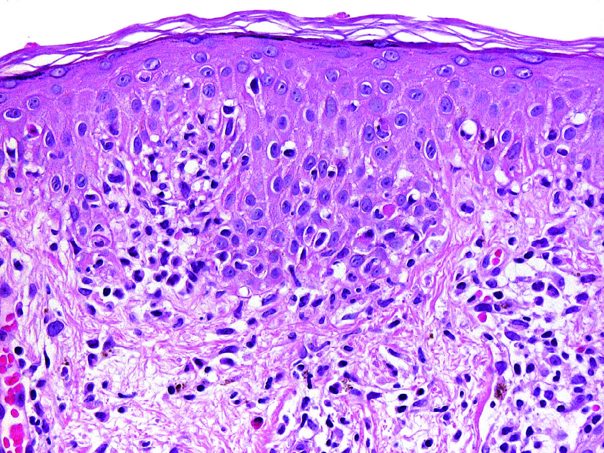
Fig.21.3 B
s.461
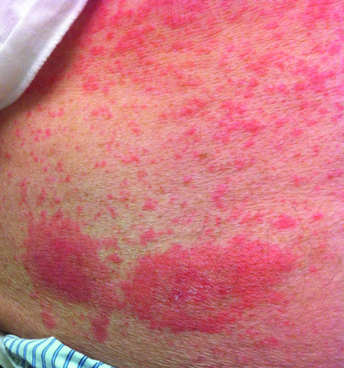
Fig.21.5 A
s.463
Fig.21.5 B
s.463
Fig.21.6
s.464
Fig.21.7 A
s.465
Fig.21.7 B
s.465
Fig.21.8
s.466
Fig.21.9
s.467
Fig.21.10 A
s.468
Fig.21.10 B
s.468
Fig.21.10 C
s.468
Fig.21.10 D
s.468
Fig.21.10 E
s.468
Fig.21.12 A
s.469
Fig.21.12 B
s.469
Fig.21.13 A
s.469
Fig.21.13 B
s.469
Fig.21.16 A
s.473
Fig.21.16 B
s.473
Fig.21.16 C
s.473
Fig.21.16 D
s.473
Fig.21.16 E
s.473
Fig.21.17
s.473
Fig.21.18 A
s.474
Fig.21.18 B
s.474
Fig.21.19
s.477
Fig.21.20 A
s.478
Fig.21.20 B
s.478
Fig.21.20 C
s.478
Fig.21.21
s.478
Fig.21.22
s.480
Fig.21.23 A
s.481
Fig.21.23 B
s.481
Fig.21.24
s.481
Fig.21.25
s.483
✕ Kapat