Bolognia 5e
·
Dermatoloji Çalışma Paneli
▤
Anasayfa
📖
Özetler
🃏
Flashcard
❓
Quiz
🖼
Atlas
◐
Ch 24
Kütanöz Vaskülit
Vol 1 · sayfa 418 · §4
← Ch 21
Ch 25 →
📖
Özet
🃏
Flashcard
❓
Quiz
🖼
Atlas
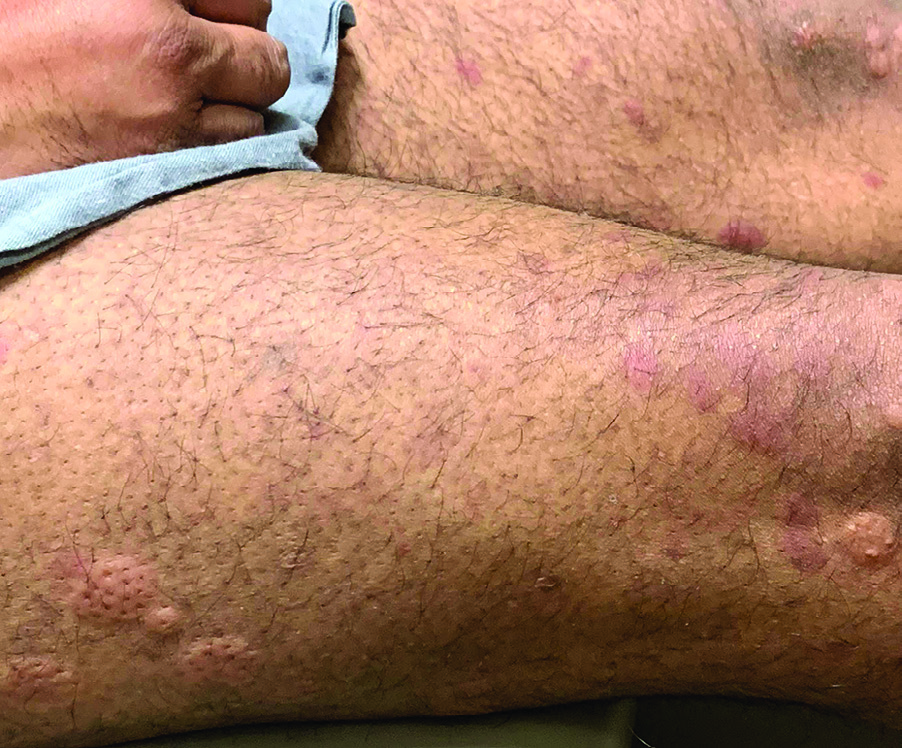
Fig.24.2 A
s.536
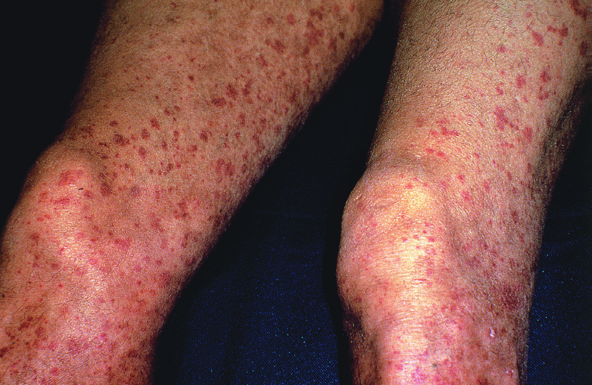
Fig.24.2 B
s.536
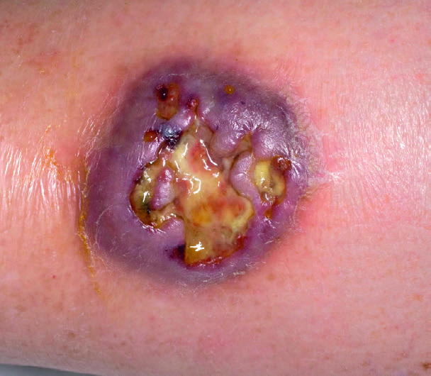
Fig.24.2 C
s.536
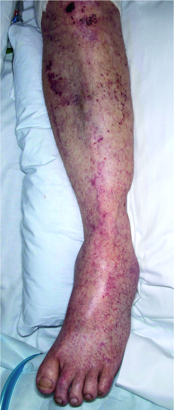
Fig.24.2 D
s.536
Fig.24.3 A
s.537
Fig.24.3 B
s.537
Fig.24.3 C
s.537
Fig.24.3 D
s.537
Fig.24.3 E
s.537
Fig.24.3 F
s.537
Fig.24.5
s.541
Fig.24.6 A
s.541
Fig.24.6 B
s.541
Fig.24.6 C
s.541
Fig.24.9 A
s.545
Fig.24.9 B
s.545
Fig.24.10 A
s.545
Fig.24.10 B
s.545
Fig.24.13 A
s.548
Fig.24.13 B
s.548
Fig.24.13 C
s.548
Fig.24.13 D
s.548
Fig.24.13 E
s.548
Fig.24.13 F
s.548
Fig.24.13 G
s.548
Fig.24.15 A
s.550
Fig.24.15 B
s.550
Fig.24.17 A
s.553
Fig.24.17 B
s.553
Fig.24.17 C
s.553
Fig.24.17 D
s.553
Fig.24.17 E
s.553
Fig.24.17 F
s.553
Fig.24.18 A
s.556
Fig.24.18 B
s.556
Fig.24.20 A
s.558
Fig.24.20 B
s.558
Fig.24.21 A
s.560
Fig.24.21 B
s.560
Fig.24.21 C
s.560
Fig.24.21 D
s.560
Fig.24.21 E
s.560
Fig.24.22
s.560
Fig.24.23
s.561
✕ Kapat