Bolognia 5e
·
Dermatoloji Çalışma Paneli
▤
Anasayfa
📖
Özetler
🃏
Flashcard
❓
Quiz
🖼
Atlas
◐
Ch 25
Nötrofilik Dermatozlar
Vol 1 · sayfa 450 · §4
← Ch 24
Ch 29 →
📖
Özet
🃏
Flashcard
❓
Quiz
🖼
Atlas
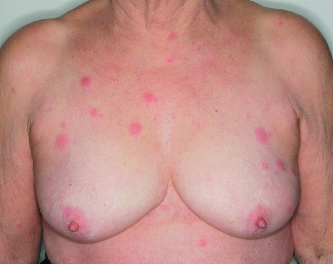
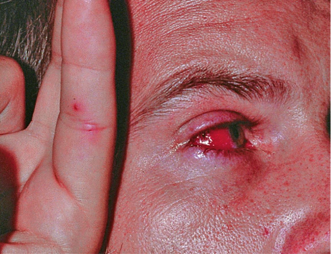
Fig.25.2 A
s.574
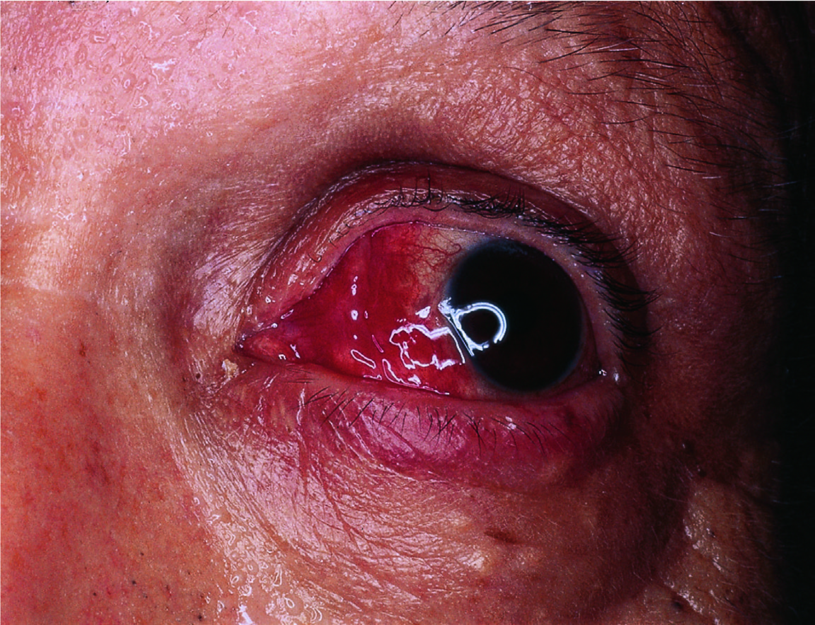
Fig.25.2 B
s.574
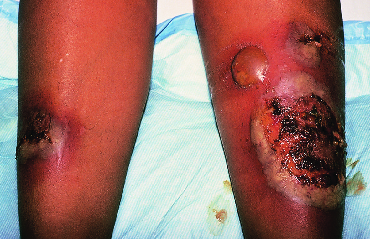
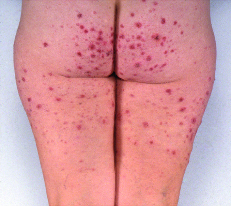
Fig.25.3 A
s.575
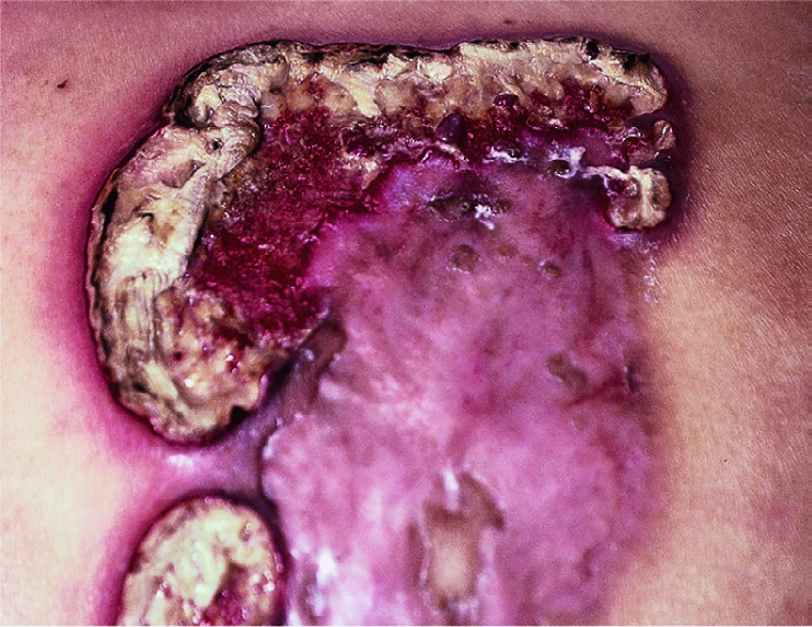
Fig.25.3 B
s.575
Fig.25.3 C
s.575
Fig.25.4
s.575
Fig.25.7 A
s.577
Fig.25.7 B
s.577
Fig.25.9 A
s.579
Fig.25.9 B
s.579
Fig.25.10 A
s.579
Fig.25.10 B
s.579
Fig.25.10 C
s.579
Fig.25.11 A
s.580
Fig.25.11 B
s.580
Fig.25.12
s.580
Fig.25.13
s.580
Fig.25.14
s.581
Fig.25.15 A
s.584
Fig.25.15 B
s.584
Fig.25.16
s.585
Fig.25.17 A
s.588
Fig.25.17 B
s.588
✕ Kapat