Bolognia 5e
·
Dermatoloji Çalışma Paneli
▤
Anasayfa
📖
Özetler
🃏
Flashcard
❓
Quiz
🖼
Atlas
◐
Ch 36
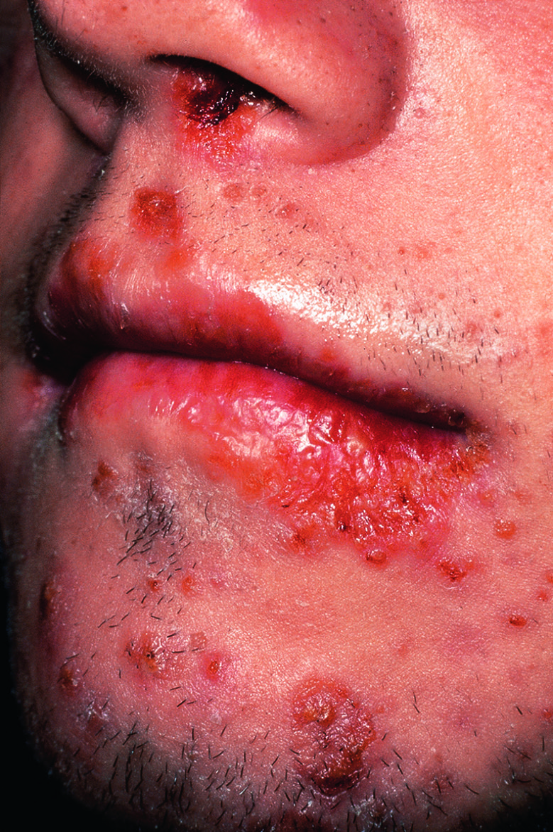
Akne Vulgaris
Vol 1 · sayfa 592 · §6
← Ch 30
Ch 37 →
📖
Özet
🃏
Flashcard
❓
Quiz
🖼
Atlas
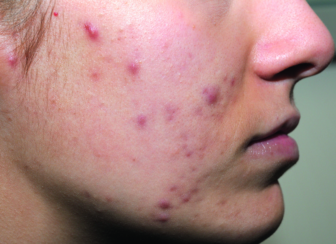
Fig.36.2 A
s.748
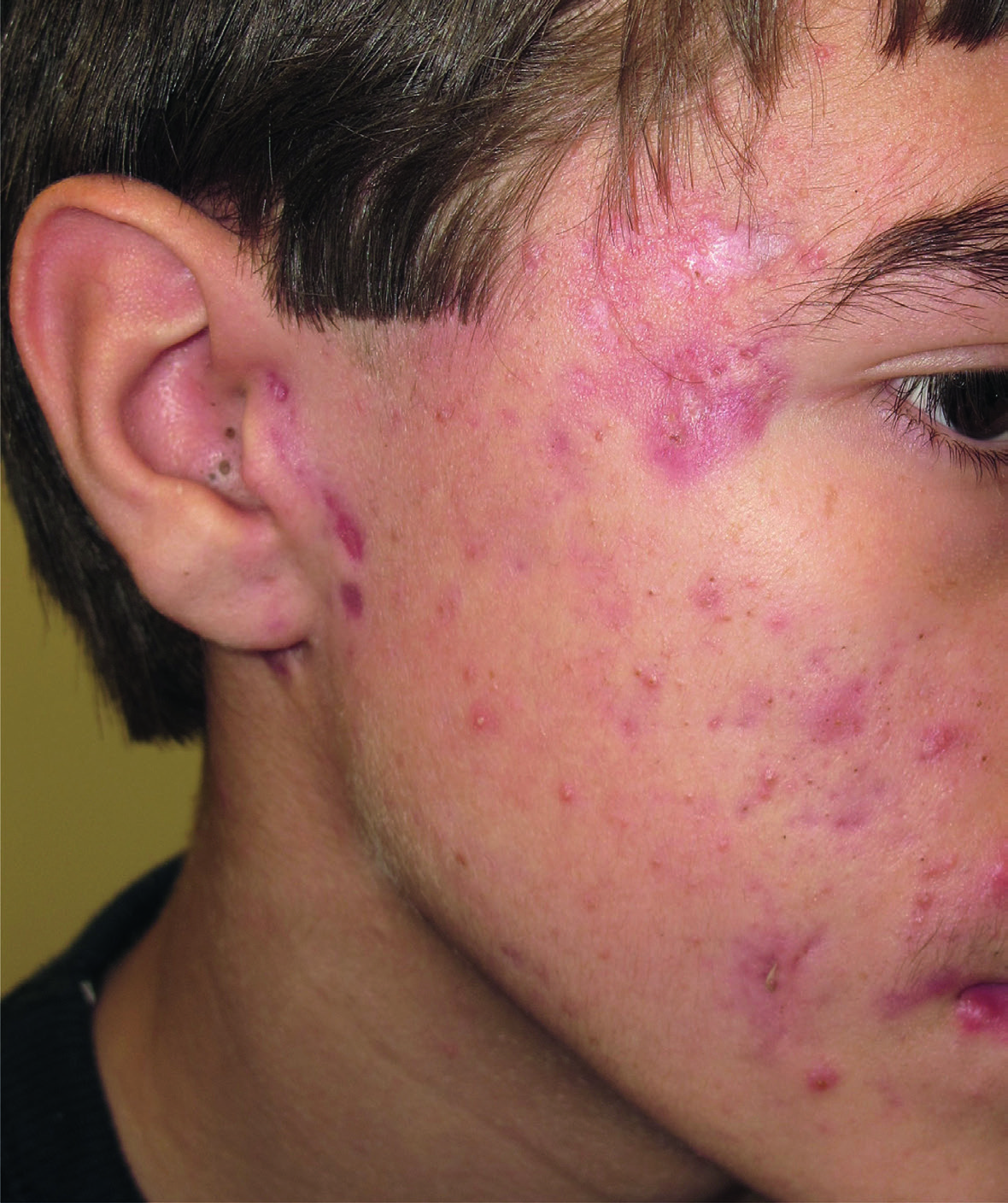
Fig.36.2 B
s.748
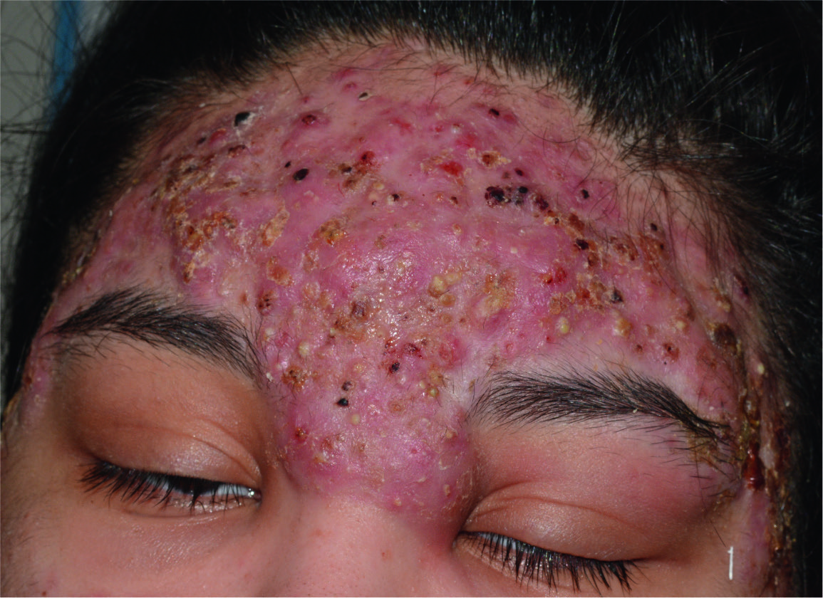
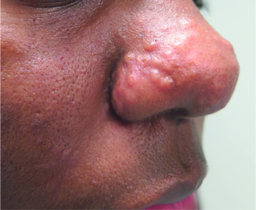
Fig.36.3
s.748
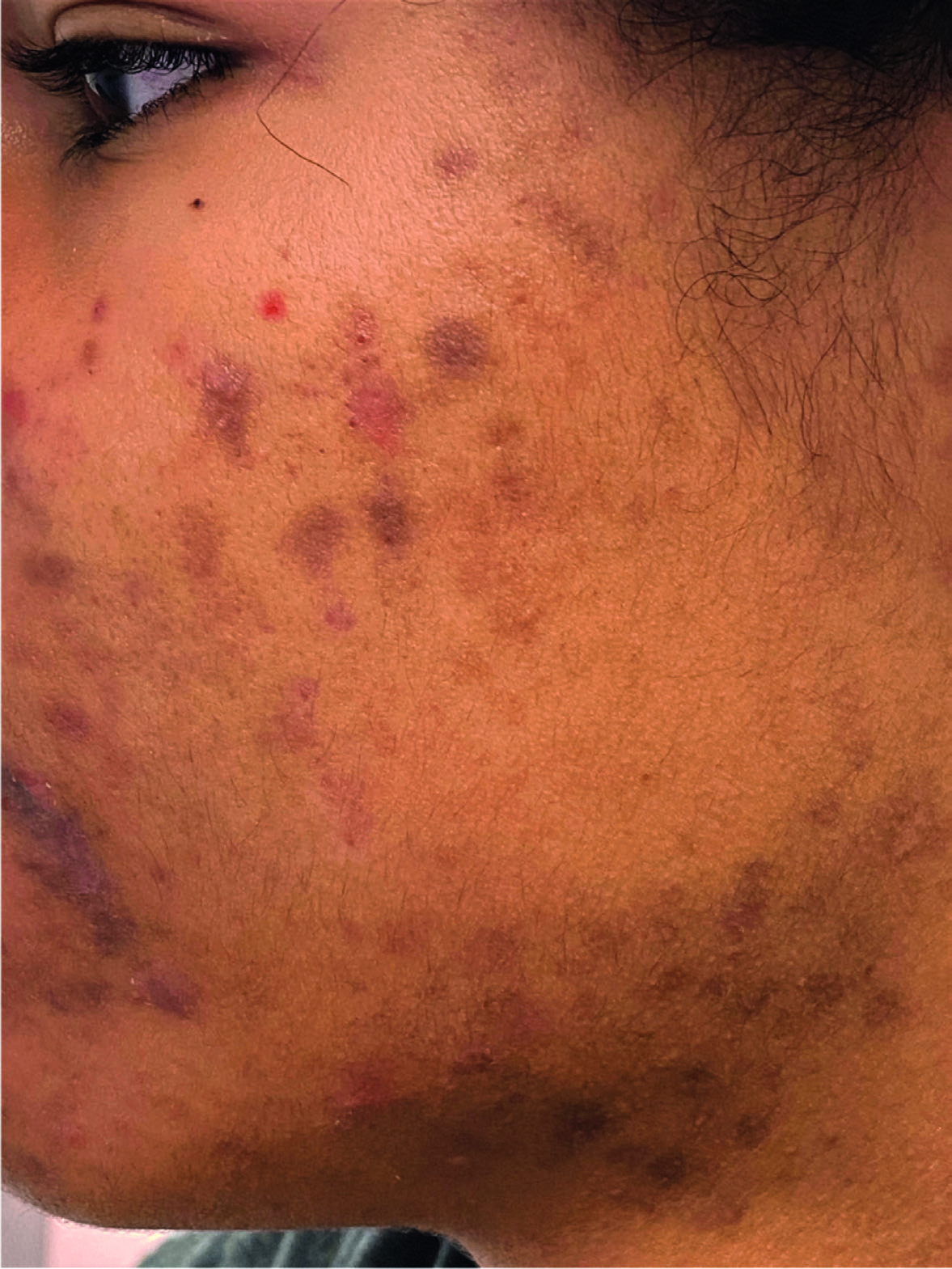
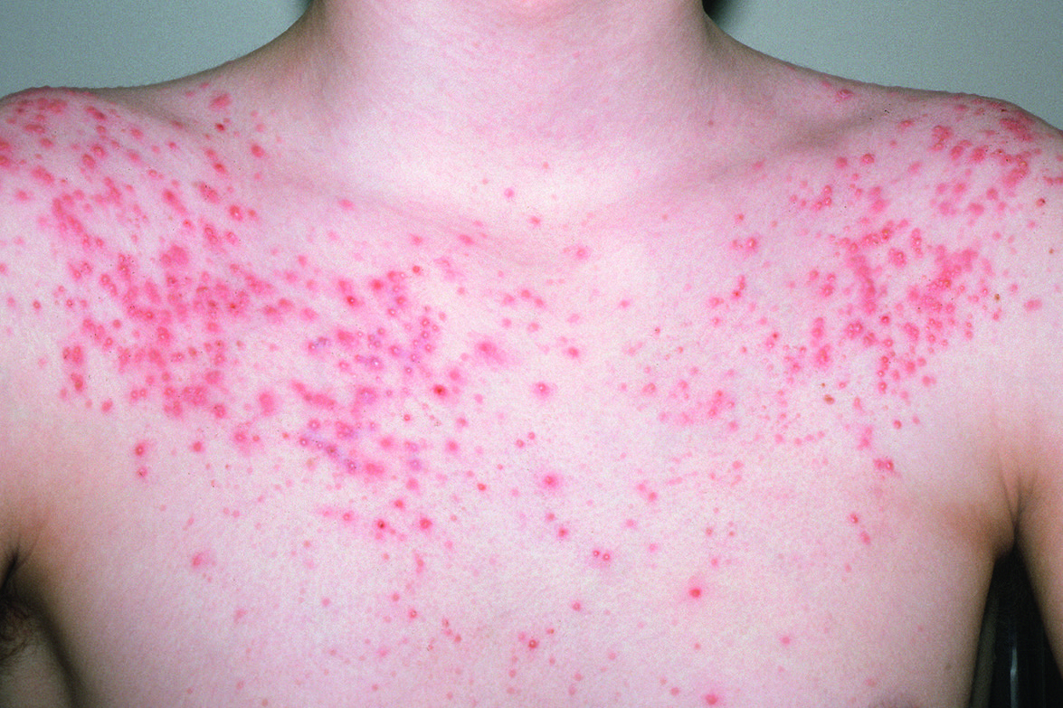
Fig.36.4
s.749
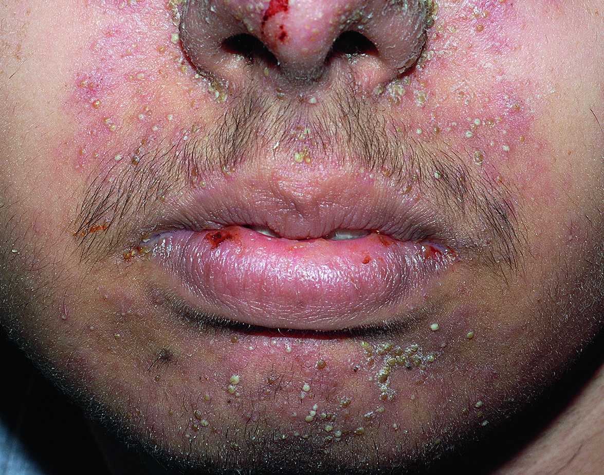
Fig.36.5
s.749
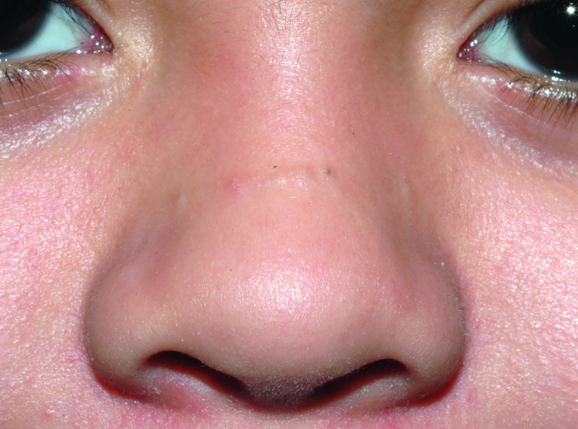
Fig.36.6
s.750
Fig.36.7
s.750
Fig.36.8 A
s.751
Fig.36.8 B
s.751
Fig.36.10 A
s.751
Fig.36.10 B
s.751
Fig.36.11
s.751
Fig.36.12 A
s.752
Fig.36.12 B
s.752
Fig.36.13
s.753
Fig.36.14
s.753
Fig.36.15 A
s.754
Fig.36.15 B
s.754
Fig.36.15 C
s.754
Fig.36.15 D
s.754
Fig.36.17 A
s.760
Fig.36.17 B
s.760
✕ Kapat