Bolognia 5e
·
Dermatoloji Çalışma Paneli
▤
Anasayfa
📖
Özetler
🃏
Flashcard
❓
Quiz
🖼
Atlas
◐
Ch 71
Tırnak Hastalıkları
Vol 1 · sayfa 1214 · §11
← Ch 69
Ch 74 →
📖
Özet
🃏
Flashcard
❓
Quiz
🖼
Atlas
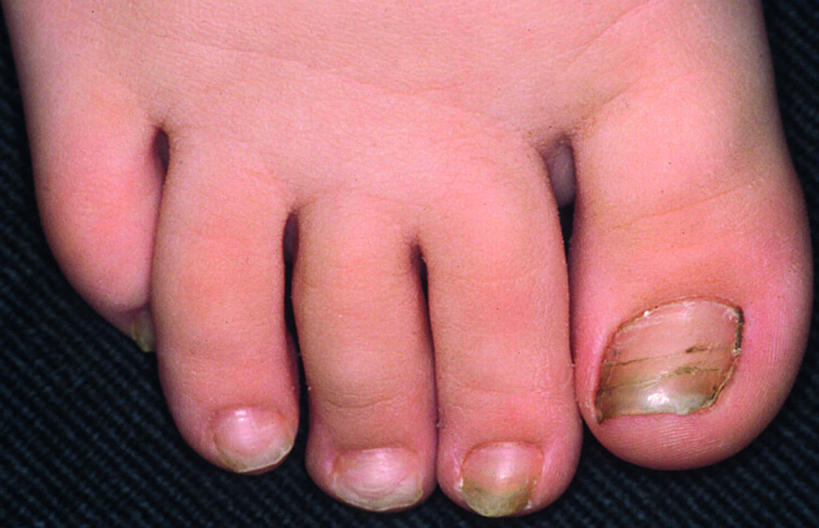
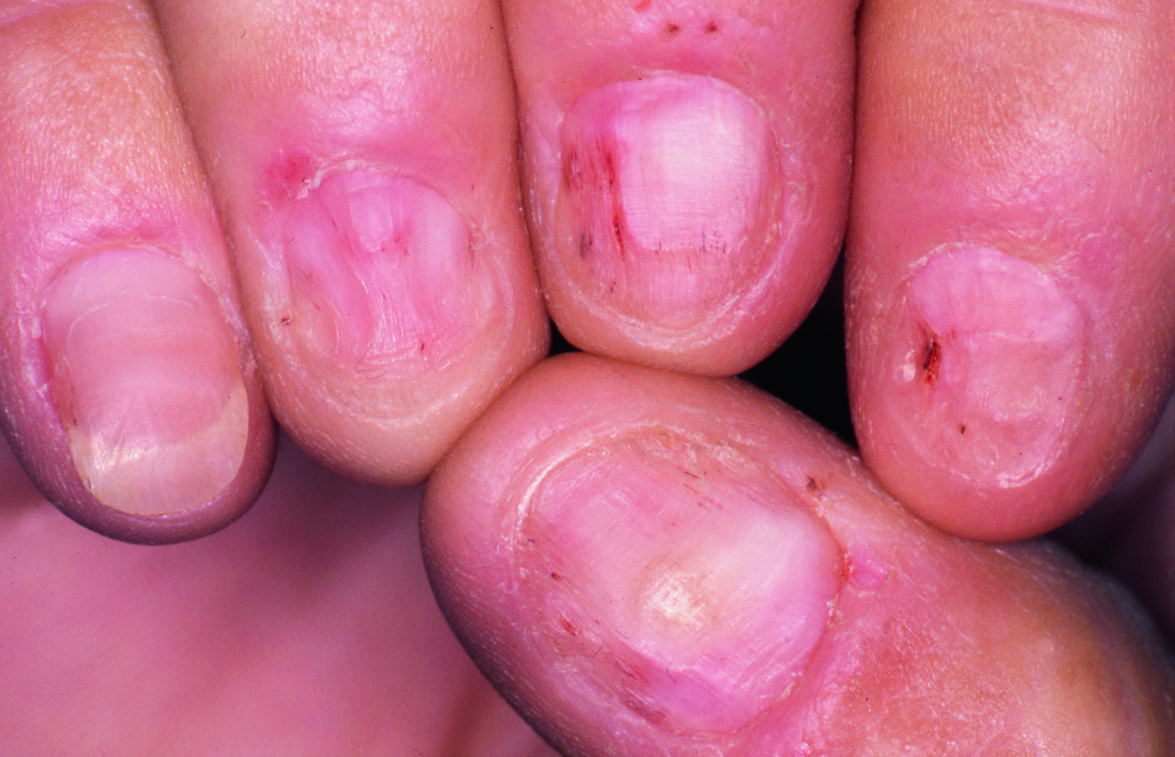
Fig.71.3 A
s.1472
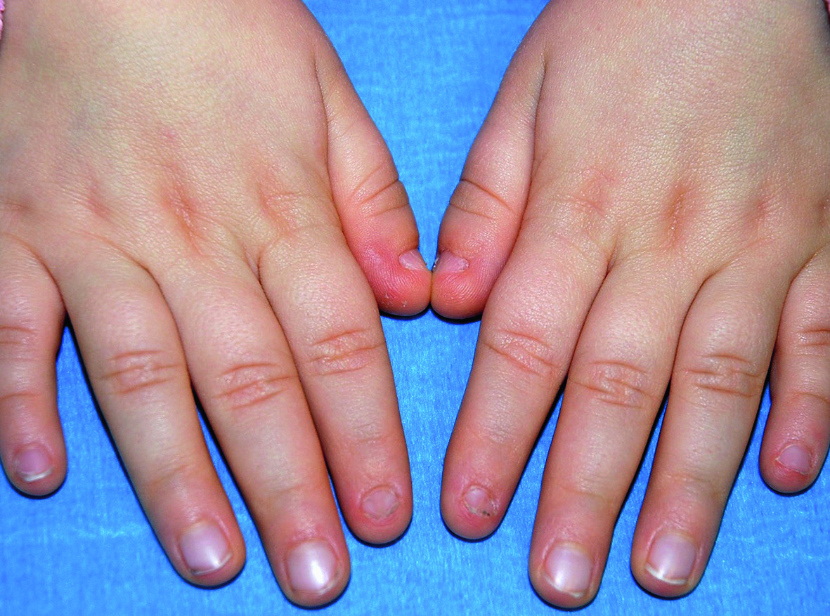
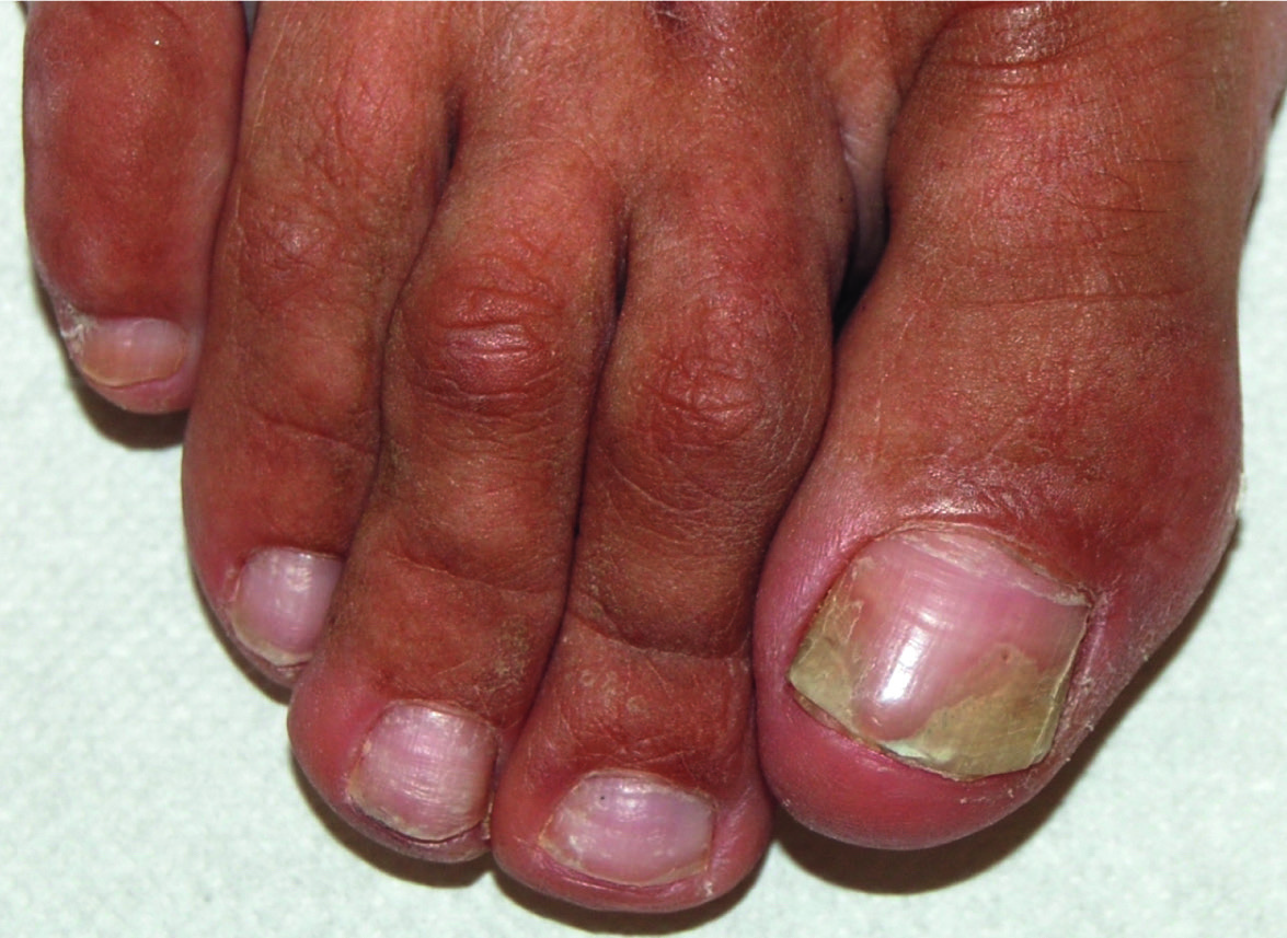
Fig.71.3 B
s.1473
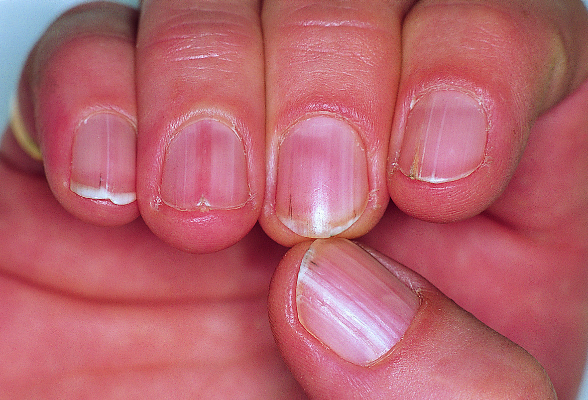
Fig.71.4
s.1473
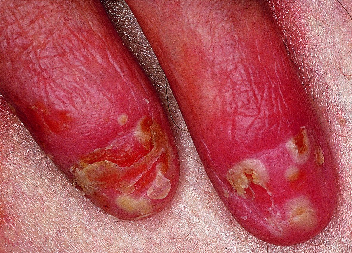
Fig.71.5
s.1473
Fig.71.6
s.1474
Fig.71.8 A
s.1476
Fig.71.8 B
s.1476
Fig.71.10
s.1477
Fig.71.11
s.1478
Fig.71.12
s.1478
Fig.71.13
s.1479
Fig.71.14
s.1479
Fig.71.15
s.1480
Fig.71.16
s.1481
Fig.71.17
s.1481
Fig.71.18
s.1482
Fig.71.19
s.1483
Fig.71.20 A
s.1484
Fig.71.20 B
s.1484
Fig.71.21
s.1484
Fig.71.22
s.1485
Fig.71.23 A
s.1486
Fig.71.23 B
s.1486
Fig.71.24 A
s.1486
Fig.71.24 B
s.1486
Fig.71.25 A
s.1486
Fig.71.25 B
s.1486
Fig.71.26 A
s.1486
Fig.71.26 B
s.1486
Fig.71.27
s.1487
Fig.71.28
s.1487
✕ Kapat